. 2005 Jan;25(2):706-15.
doi: 10.1128/MCB.25.2.706-715.2005.

Interaction between GATA and the C/EBP family of transcription factors is critical in GATA-mediated suppression of adipocyte differentiation

Affiliations

Interaction between GATA and the C/EBP family of transcription factors is critical in GATA-mediated suppression of adipocyte differentiation

Qiang Tong et al. Mol Cell Biol. 2005 Jan.

Abstract

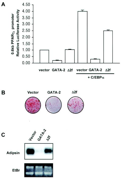
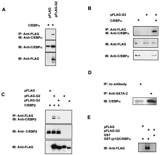
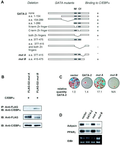
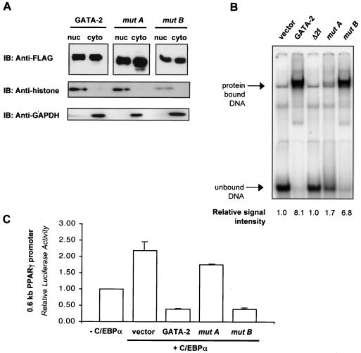
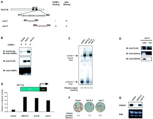
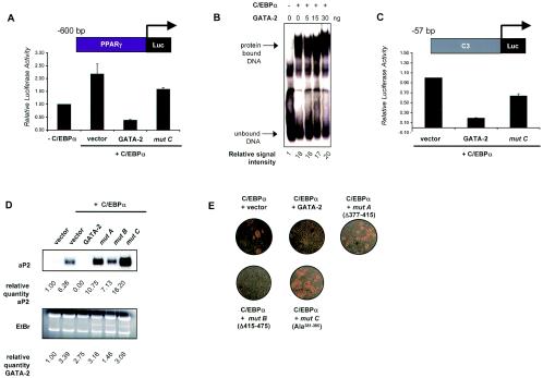
We have previously demonstrated that GATA-2 and GATA-3 are expressed in adipocyte precursors and control the preadipocyte-to-adipocyte transition. Constitutive expression of both GATA-2 and GATA-3 suppressed adipocyte differentiation, partially through direct binding to the peroxisome proliferator-activated receptor gamma (PPARgamma) promoter and suppression of its basal activity. In the present study, we demonstrate that both GATA-2 and GATA-3 form protein complexes with CCAAT/enhancer binding protein alpha (C/EBPalpha) and C/EBPbeta, members of a family of transcription factors that are integral to adipogenesis. We mapped this interaction to the basic leucine zipper domain of C/EBPalpha and a region adjacent to the carboxyl zinc finger of GATA-2. The interaction between GATA and C/EBP factors is critical for the ability of GATA to suppress adipocyte differentiation. Thus, these results show that in addition to its previously recognized function in suppressing PPARgamma transcriptional activity, interaction of GATA factors with C/EBP is necessary for their ability to negatively regulate adipogenesis.


Figures

FIG. 1.
FIG. 1.
GATA-mediated suppression of adipogenesis is not fully reversed by disruption of DNA binding. Renilla luciferase vector was included as an internal control to account for transfection efficiency. (A) Relative luciferase activity of a PPARγ promoter construct (PPARγ2) driving a luciferase reporter was examined in NIH 3T3 cells expressing GATA-2 or a GATA-2 mutant in which both zinc fingers have been deleted (Δ2f) in the presence (+) or absence of C/EBPα. The values reported here represent the means ± standard errors of the means (SEMs) of relative luciferase activities. Error bars indicate SEMs. (B) Oil red O staining of 3T3-F442A cells constitutively expressing either wild-type GATA-2 or a mutant lacking both zinc fingers (Δ2f) and stimulated for adipocyte differentiation for 8 days with insulin. (C) Northern blot analysis for gene expression of adipsin, which is expressed in a differentiation-dependent manner, in differentiated 3T3-F442A cells. Ethidium bromide (EtBr) staining demonstrates equal loading in each lane as well as the integrity of mRNA.
FIG. 2.
FIG. 2.
Interaction of GATA-2 or GATA-3 with C/EBPα or C/EBPβ. (A) COS-7 cells were transfected with expression plasmids for C/EBPα along with FLAG-tagged GATA-2 or pFLAG vector control. Cell lysates were harvested and immunoprecipitated (IP) with an anti-FLAG antibody conjugated to agarose beads, and the proper expression of C/EBPα was detected by immunoblotting (IB) with an antibody against C/EBPα. The expression of GATA-2 or C/EBPα was confirmed by direct immunoblot analysis of 10 μg of whole-cell lysate with anti-FLAG or anti-C/EBPα antibodies, respectively. (B) C/EBPα was also detected in anti-FLAG immunoprecipitants of cells expressing FLAG-tagged GATA-3 (pFLAG-G3) and C/EBPα. Coprecipitation analysis was performed as described for panel A. (C) Detection of C/EBPβ protein with an isoform-specific anti-C/EBPβ antibody in anti-FLAG immunoprecipitants of COS-7 cells expressing C/EBPβ along with either FLAG-tagged GATA-2 or GATA-3. Coprecipitation analysis was performed as described for panel A. (D) Detection of CEBP-GATA interaction in intact cells. 3T3-L1 cells were stimulated to differentiate for 24 h, at which time whole-cell lysates were collected and immunoprecipitated with anti-GATA-2 antibody, followed by immunoblot analysis with an anti-C/EBPα antibody. Immunoprecipitation of lysates with no antibody controlled for nonspecific binding of protein complexes to protein A-Sepharose beads. (E) A 12-kDa carboxy-terminal fragment of C/EBPα was expressed as a fusion protein with GST [GST-(p12)C/EBPα] and affinity purified with glutathione-Sepharose. Bound GST-(p12)C/EBPα recombinant proteins or GST alone were incubated with cell lysates of COS-7 cells expressing FLAG-tagged GATA-2. The presence of GATA-2 bound to C/EBPα was detected by immunoblot analysis with an anti-FLAG antibody.
FIG. 3.
FIG. 3.
Characterization of GATA-2 and C/EBP interaction in adipogenesis. (A) Schematic diagram of various GATA deletion mutants and their ability to bind C/EBPα, as evaluated by coimmunoprecipitation analyses as described in the legend to Fig. 2A. Nf and Cf, amino- and carboxy-terminal zinc fingers, respectively. N-term, N-terminal; C-term, C-terminal. (B) A DNA construct for either FLAG-tagged mutant A or B was transfected into COS-7 cells along with an expression plasmid for C/EBPα. The association of C/EBPα with these GATA mutants was detected by immunoprecipitation (IP) with anti-FLAG antibody-conjugated agarose beads, followed by immunoblotting (IB) with an anti-C/EBPα antibody. The proper expression of C/EBPα and GATA-2 mutants in this protein complex was confirmed by immunoblot analysis using anti-C/EBPα and anti-FLAG antibodies, respectively. (C) Oil red O staining of differentiated 3T3-F442A cells constitutively expressing wild-type GATA-2 or GATA-2 mutant A or B. Expression levels of GATA-2 and the mutants were confirmed by quantitative RT-PCR and numerically expressed as a difference (n-fold) of the level of the vector control. N/A, not analyzed. (D) Northern blot analysis of expression of adipogenic genes adipsin and PPARγ in differentiated 3T3-F442A cells constitutively expressing wild-type GATA-2, mutant A, or mutant B. Ethidium bromide (EtBr) staining was included to demonstrate loading in each lane and the integrity of RNA.
FIG. 4.
FIG. 4.
Analysis of nuclear localization and DNA binding properties of mutant A [GATA-2 (Δ377-415)] and mutant B [GATA-2 (Δ415-475)]. (A) Cytosolic (cyto) and nuclear (nuc) fractions were prepared from cell lysates of COS-7 cells expressing FLAG-tagged GATA-2, mutant A, or mutant B. The presence of GATA-2 in these subcellular fractions was detected by immunoblotting (IB) with an anti-FLAG antibody. The nuclear fraction was confirmed by detection of the nuclear protein histone, and the cytosolic fraction was confirmed by detection of the housekeeping protein GAPDH. (B) Nuclear extracts of COS-7 cells expressing either GATA-2, GATA-2 lacking both zinc fingers (Δ2f), or mutant A or B were analyzed by electrophoretic mobility shift assay with an oligonucleotide consisting of consensus GATA binding sites (upper panel). The signals are quantitated and expressed numerically under each lane. (C) C/EBPα-activated promoter activity of PPARγ was examined in the presence of wild-type GATA-2 or deletion mutants A or B. The values reported here represent the mean ± SEM relative luciferase activity. Renilla luciferase activity was used as an internal control.
FIG. 5.
FIG. 5.
Detailed mapping and functional analysis of GATA-C/EBP interaction. (A) Schematic diagram of specific amino acid substitutions generated in mutant C (aa 381 to 385) and mutant D (aa 388 to 394). Nf and Cf, amino- and carboxy-terminal zinc fingers, respectively. The ability of each mutant to interact with C/EBPα or to bind to target DNA has been summarized in the column next to the diagram. (B) Anti-C/EBPα antibody was used to detect C/EBPα in protein complexes immunoprecipitated (IP) with an anti-FLAG antibody from lysates of COS-7 cells expressing FLAG-tagged mutant C or mutant D along with C/EBPα. (C) An electrophoretic mobility shift assay was used to test the ability of mutant C to bind a cognate DNA sequence. Nuclear extracts from COS-7 cells expressing GATA-2, mutant A, or mutant C were used. Binding is quantitated as described in the legend to Fig. 3 and expressed numerically under each lane. (D) Cytosolic and nuclear fractions were prepared from cell lysates of COS-7 cells expressing mutant C. The presence of GATA-2 in these subcellular fractions was detected by immunoblotting (IB) with an anti-FLAG antibody. The nuclear fraction was confirmed by detection of the nuclear protein histone, and the cytosolic fraction was confirmed by detection of the housekeeping protein GAPDH. (E) GATA-mediated activation of the interleukin-5 luciferase reporter was examined in the presence of wild-type GATA-2, mutant B, or mutant C. (F) Oil red O staining of differentiated 3T3-F442A cells constitutively expressing GATA-2 or mutant C. Expression of GATA-2 and mutants were confirmed by quantitative RT-PCR and numerically expressed as described in the legend to Fig. 4. (G) Northern blot analysis of adipsin expression in differentiated 3T3-F442A cells constitutively expressing GATA-2 or mutant C. EtBr, ethidium bromide.
FIG. 6.
FIG. 6.
Effect of various GATA-2 mutants on C/EBPα-activated adipogenesis in NIH 3T3 cells. (A) C/EBPα-activated promoter activity of PPARγ was examined in the presence of wild-type GATA-2 or mutant C. (B) Electrophoretic mobility shift assay was used to test the ability of C/EBPα to bind a cognate DNA sequence in the presence of increasing amounts of GATA-2. Nuclear extracts from COS-7 cells expressing C/EBPα were used along with various amounts of recombinant GATA-2. (C) C/EBPα-activated promoter activity of C3Luc in the presence of wild-type GATA-2 or mutant C. For panels A and C, the values reported represent the mean luciferase activity values ± SEM. Renilla luciferase activity was used as an internal control. (D) Northern blot analysis of total RNA from the same cells was used to detect the expression of the late adipogenic marker aP2. Ethidium bromide (EtBr) staining was included to demonstrate equal loading in each lane as well as the integrity of RNA. Signal intensities of aP2 were calculated using Personal FX phosphorimaging software (Bio-Rad). Exogenous expression of GATA-2 and mutants were confirmed by quantitative RT-PCR. Both values are numerically expressed as a difference (n-fold) of the value for the vector control. (E) Phase-contrast micrographs of differentiated NIH 3T3 cells constitutively expressing C/EBPα along with various GATA-2 mutants.

References

    1. Blobel, G. A., T. Nakajima, R. Eckner, M. Montminy, and S. H. Orkin. 1998. CREB-binding protein cooperates with transcription factor GATA-1 and is required for erythroid differentiation. Proc. Natl. Acad. Sci. USA 95:2061-2066. - PMC - PubMed
    1. Boyes, J., P. Byfield, Y. Nakatani, and V. Ogryzko. 1998. Regulation of activity of the transcription factor GATA-1 by acetylation. Nature 396:594-598. - PubMed
    1. Cao, Z., R. M. Umek, and S. L. McKnight. 1991. Regulated expression of three C/EBP isoforms during adipose conversion of 3T3-L1 cells. Genes Dev. 5:1538-1552. - PubMed
    1. Chen, P. L., D. J. Riley, Y. Chen, and W. H. Lee. 1996. Retinoblastoma protein positively regulates terminal adipocyte differentiation through direct interaction with C/EBPs. Genes Dev. 10:2794-2804. - PubMed
    1. Crawford, S. E., C. Qi, P. Misra, V. Stellmach, M. S. Rao, J. D. Engel, Y. Zhu, and J. K. Reddy. 2002. Defects of the heart, eye, and megakaryocytes in peroxisome proliferator activator receptor-binding protein (PBP) null embryos implicate GATA family of transcription factors. J. Biol. Chem. 277:3585-3592. - PubMed

Publication types

MeSH terms

LinkOut - more resources