. 2005 Feb;6(2):125-31.
doi: 10.1631/jzus.2005.B0125.

Cytotoxicity of epigallocatechin-3-gallate to LNCaP cells in the presence of Cu2+

Affiliations

Cytotoxicity of epigallocatechin-3-gallate to LNCaP cells in the presence of Cu2+

Hai-ning Yu et al. J Zhejiang Univ Sci B. 2005 Feb.

Abstract

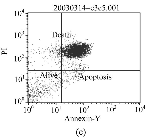
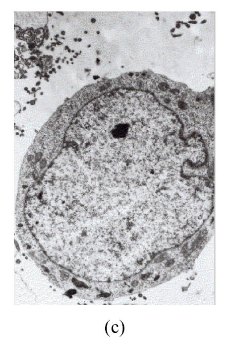
Epigallocatechin-3-gallate (EGCG) has shown remarkably anti-cancer activity, with its bioactivity being related to reactive conditions, such as pH and metal ions. The present study investigated the degradation of EGCG and its effect on prostate cancer cell in the presence of Cu2+. EGCG was incubated with prostate cancer cells, LNCaP, pretreated with or without Cu2+. EGCG in F-12 medium was quantified using HPLC and the viability of cells was assessed by gel electrophoresis, flow cytometry, and electron microscope. The results of HPLC showed that EGCG degraded completely within 12 h in F-12 medium with or without Cu2+. Gel electrophoresis and flow cytometry did not detect apoptosis of LNCaP cells when they were incubated with EGCG. Electron microscopy examination revealed that EGCG-Cu2+ complex led to damage of cytoplasm membrane in LNCaP cells. It was speculated that not EGCG, but its oxide and complex with Cu2+, are the bioactive components responsible for its cytotoxicity to LNCaP prostate cancer cells.


Figures

Fig. 1
Fig. 1
HPLC chromatogram of F-12 with LNCaP cells incubated with 320 μmol/L CuSO4 pretreated with 200 μmol/L EGCG for 24 h
Fig. 2
Fig. 2
Gel electrophoretic analysis of DNA from LNCaP cells treated with EGCG and CuSO4 a: LNCaP cells; b: LNCaP cells treated with 32 μmol/L CuSO4 before 150 μmol/L EGCG was added; c: LNCaP cells incubated with 32 μmol/L CuSO4 after 150 μmol/L EGCG was added; d: LNCaP cells treated with 200 μmol/L EGCG after 320 μmo/L CuSO4 was added; e: LNCaP cells incubated with 320 μmol/L CuSO4 after 200 μmol/L EGCG was added
Fig. 3
Fig. 3
Detection of apoptosis by flow cytometry (partial figures are shown, all data are shown in Table 2). (a) Normal LNCaP cells; (b) LNCaP cellss treated with 10 μmol/L EGCG; (c) LNCaP cells incubated with 200 μmol/L EGCG after 320 μmol/L CuSO4 was added
Fig. 3
Fig. 3
Detection of apoptosis by flow cytometry (partial figures are shown, all data are shown in Table 2). (a) Normal LNCaP cells; (b) LNCaP cellss treated with 10 μmol/L EGCG; (c) LNCaP cells incubated with 200 μmol/L EGCG after 320 μmol/L CuSO4 was added
Fig. 3
Fig. 3
Detection of apoptosis by flow cytometry (partial figures are shown, all data are shown in Table 2). (a) Normal LNCaP cells; (b) LNCaP cellss treated with 10 μmol/L EGCG; (c) LNCaP cells incubated with 200 μmol/L EGCG after 320 μmol/L CuSO4 was added
Fig. 4
Fig. 4
Configuration of LNCaP cells treated with different concentrations of EGCG and CuSO4. (a) Normal LNCaP cells; (b) Treated with 10 μmol/L EGCG; (c) Treated with 0.0064 μmol/L CuSO4; (d): cytoplasm membrane of LNCaP cells treated with 0.0064 μmol/L CuSO4; (e) Treated with 150 μmol/L EGCG in the presence of 32 μmol/L CuSO4; (f) Put into 150 μmol/L EGCG, then added 32 μmol/L CuSO4; (g) Before addition of 200 μmol/L EGCG, put into 320 μmol/L CuSO4; (h) Treated with 320 μmol/L CuSO4 after 200 μmol/L EGCG was added
Fig. 4
Fig. 4
Configuration of LNCaP cells treated with different concentrations of EGCG and CuSO4. (a) Normal LNCaP cells; (b) Treated with 10 μmol/L EGCG; (c) Treated with 0.0064 μmol/L CuSO4; (d): cytoplasm membrane of LNCaP cells treated with 0.0064 μmol/L CuSO4; (e) Treated with 150 μmol/L EGCG in the presence of 32 μmol/L CuSO4; (f) Put into 150 μmol/L EGCG, then added 32 μmol/L CuSO4; (g) Before addition of 200 μmol/L EGCG, put into 320 μmol/L CuSO4; (h) Treated with 320 μmol/L CuSO4 after 200 μmol/L EGCG was added

Similar articles

Cited by

References

    1. Chung LY, Cheung TC, Kong SK, Fung KP, Choy YM, Chan ZY, Kwok TT. Induction of apoptosis by green tea catechins in human prostate cancer DU145 cells. Life Sciences. 2001;68(10):1207–1214. - PubMed
    1. Chung JY, Park JO, Phyu H, Dong ZG, Yang CS. Mechanisms of inhibition of the Ras-MAP kinase signaling pathway in 30.7b Ras 12 cells by tea polyphenols (-)-epigallocatechin-3-gallate and theaflavin-3,3′-digallate. The FASEB Journal. 2001;15:2022–2024. - PubMed
    1. Cutter H, Wu LY, Kim CD, Morre J, Morre DM. Is the cancer protective effect correlated with growth inhibitions by green tea (-)-epigallocatechin gallate mediated through an antioxidant mechanism? Cancer Lett. 2001;162(2):149–154. - PubMed
    1. Gupta S, Ahmad N, Nieminent AL, Mukhtar H. Growth inhibition, cell-cycle dysregulation, and induction of apoptosis by green tea constituent (-)-epigallocatechin-3-gallate in androgen-sensitive and androgen-insensitive human prostate carcinoma cells. Toxicology and Applied Pharmacology. 2000;164(1):82–90. - PubMed
    1. Gupta S, Hussain T, Mukhtar H. Molecular pathway for (-)-epigallocatechin-3-gallate-induced cell cycle arrest and apoptosis of human prostate carcinoma cells. Archives of Biochemistry and Biophysics. 2003;410(1):177–185. - PubMed

Publication types

MeSH terms