. 2005 Jan 15;385(Pt 2):595-603.
doi: 10.1042/BJ20040971.

Apo2L/TRAIL induction and nuclear translocation of inositol hexakisphosphate kinase 2 during IFN-beta-induced apoptosis in ovarian carcinoma

Affiliations

Apo2L/TRAIL induction and nuclear translocation of inositol hexakisphosphate kinase 2 during IFN-beta-induced apoptosis in ovarian carcinoma

Bei H Morrison et al. Biochem J. .

Abstract

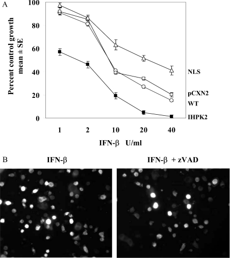
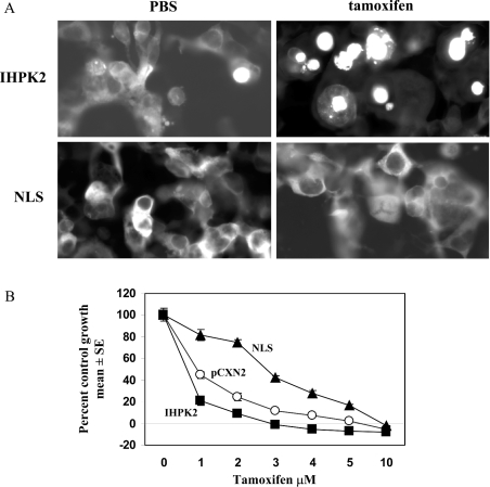
Previously, we have reported that overexpression of IHPK2 (inositol hexakisphosphate kinase 2) sensitized NIH-OVCAR-3 ovarian carcinoma cell lines to the growth-suppressive and apoptotic effects of IFN-beta (interferon-beta) treatment and gamma-irradiation. In the present study, we demonstrate that Apo2L/TRAIL (Apo2L/tumour-necrosis-factor-related apoptosis-inducing ligand) is a critical mediator of IFN-induced apoptosis in these cells. Compared with IFN-alpha2, IFN-beta is a more potent inducer of Apo2L/TRAIL and IHPK2 activity. Overexpression of IHPK2 converts IFN-alpha2-resistant cells into cells that readily undergo apoptosis in response to IFN-alpha2. In untreated cells transfected with IHPK2-eGFP (where eGFP stands for enhanced green fluorescent protein), the fusion protein is localized to the cytoplasm and perinuclear region. After treatment with IFN-beta, IHPK2-eGFP translocated to the nucleus. In cells transfected with mutant IHPK2-NLS-eGFP (where NLS stands for nuclear localization sequence), containing point mutations in the NLS, the fusion protein remained trapped in the cytoplasm, even after IFN-beta treatment. Cells expressing mutant NLS mutation were more resistant to IFN-beta. The IC50 value of IHPK2-expressing cells was 2-3-fold lower than vector control. The IC50 value of NLS-mutant-expressing cells was 3-fold higher than vector control. Blocking antibodies to Apo2L/TRAIL or transfection with a dominant negative Apo2L/TRAIL receptor (DR5Delta) inhibited the antiproliferative effects of IFN-beta. Thus overexpression of IHPK2 enhanced apoptotic effects of IFN-beta, and expression of the NLS mutant conferred resistance to IFN-beta. Apo2L/TRAIL expression and nuclear localization of IHPK2 are both required for the induction of apoptosis by IFN-beta in ovarian carcinoma.


Figures

Figure 1
Figure 1. Antiproliferative effects of IFNs in NIH-OVCAR-3 cells expressing IHPK2
Upper panel, antiproliferative effects of IFN-β and -α in ovarian carcinoma cells. NIH-OVCAR-3 cells were stably transfected with vector alone (pCXN2) and grown in the presence of 5–1000 units/ml IFN-β (black bars) or IFN-α2 (white bars). After 7 days, cells were fixed and stained with sulphorhodamine B. A570 of bound dye was measured and expressed as percentage of untreated controls. Each data point represents means±S.E.M. for eight replicates. Negative values on y-axis indicate death of initially plated cells. Lower panel, NIH-OVCAR-3 cells were stably transfected with IHPK2 cDNA ligated into the pCXN2 mammalian expression vector. Cells were grown in the presence of IFN-β or -α as above for 7 days. Abbreviation: U, unit.
Figure 2
Figure 2. IFN-β induces apoptosis in NIH-OVCAR-3 cells
TUNEL assay: NIH-OVCAR-3 cells stably expressing pCXN2 were treated with PBS, IFN-α2 or -β (1000 units/ml) for 48 h, labelled with Br-dUTP using TdT, and stained with FITC-conjugated anti-BrdU. Percentage of FITC-positive cells was determined by flow cytometry.
Figure 3
Figure 3. (A) Induction of Apo2L/TRAIL mRNA by IFNs and (B) induction of IHPK2 protein by IFNs
(A) NIH-OVCAR-3 cells were treated for 2–16 h with IFN-α2 or -β (1000 units/ml). Total RNA (40 μg) from these cells was subjected to Northern-blot analysis. Blots were hybridized with 32P-labelled specific probes for Apo2L/TRAIL mRNA (upper) and GAP3DH mRNA (lower) as control. (B) Cells were treated with IFNs as above. Cell lysates were subjected to Western-blot analysis. The blot was probed with anti-actin mAb as control.
Figure 4
Figure 4. Effect of anti-Apo2L/TRAIL blocking antibody
NIH-OVCAR-3 cells were stably transfected with pCXN2 vector (white bars) or IHPK2 (black bars). Cells were grown in the presence of PBS, affinity-purified anti-Apo2L/TRAIL antibody (mAb), IFN-β (1000 units/ml), mAb+IFN-β, IgG (isotype control)+IFN-β, IFN-α2 (1000 units/ml), mAb+IFN-α2 or IgG+IFN-α2. TUNEL assay was performed as in Figure 2. Each data point represents means±S.E.M. for three replicates.
Figure 5
Figure 5. Inhibition of Apo2L/TRAIL receptor blocks IFN-β-induced apoptotis
Vector alone (white bars) or a truncated version of the Apo2L/TRAIL DR5 receptor that functions in a dominant negative manner (DR5Δ; black bars) was stably transfected into NIH-OVCAR-3 cells. Cells were treated with IFN-β or -α2 (1000 units/ml) for 72 h. Caspase 3 enzymic activity was determined by measuring cleavage of Ac-DEVD-AMC (a fluorogenic substrate) in cell extracts. Where indicated, extracts from IFN-treated cells (72 h) were preincubated with the caspase 3 inhibitor Ac-DEVD-CHO before the addition of substrate. Relative fluorescence of substrate control was subtracted as background emission.
Figure 6
Figure 6. Expression of endogenous IHPK2
NIH-OVCAR-3 cells grown on chamber slides were treated with PBS or IFN-β or -α2 (200 units/16 h per ml). Cells were stained with monoclonal anti-IHPK2 antibody followed with HRP–goatanti-mouse secondary antibody. The chromogen diaminobenzidine is shown in dark grey.
Figure 7
Figure 7. Expression of eGFP fusion proteins
Tagged fusion proteins were created as described in the Experimental section. NIH-OVCAR-3 cells were stably transfected with eGFP-IHPK2 or eGFP-NLS, and then treated with PBS or IFN-β (200 units/ml×16 h). Nuclei were stained blue with DAPI (4,6-diamidino-2-phenylindole) and the fusion proteins fluoresce in green. Nuclear translocation of eGFP-IHPK2 occurs during apoptosis, but eGFP-NLS remains in the cytoplasm.
Figure 8
Figure 8. (A) Effect of NLS mutation on IFN-β antiproliferative activity in ovarian carcinoma cells and (B) effect of caspase inhibitor on eGFP-IHPK2 translocation
(A) Untransfected NIH-OVCAR-3 cells (WT, wild-type), or cells stably transfected with vector alone (pCXN2), IHPK2, or the NLS mutant were grown in the presence of IFN-β for 7 days. Cells overexpressing IHPK2 were more sensitive, whereas cells expressing the NLS mutant were more resistant to IFN-β. (B) Cells transfected with eGFP-IHPK2 were exposed to IFN-β (200 units/ml) and either PBS or Z-VAD (300 μM), a general caspase inhibitor for 16 h. The fusion protein fluorescence is shown in white.
Figure 9
Figure 9. (A) Localization of IHPK2 in HEK-293T cells and (B) effect of NLS mutation on tamoxifen antiproliferative activity in HEK-293T cells
(A) HEK-293T cells were transfected with the eGFP tagged constructs as in Figure 7. The fusion proteins fluoresce white. Tamoxifen (30 μM, 24 h) was used to induce apoptosis. (B) Cells were stably transfected with vector alone (pCXN2), IHPK2, or the NLS mutant and grown in the presence of tamoxifen for 7 days. Cells overexpressing IHPK2 were more sensitive, whereas cells expressing the NLS mutant were more resistant to tamoxifen.

References

    1. Morrison B. H., Bauer J. A., Hu J., Grane R. W., Ozdemir A., Chawla-Sarkar M., Gong B., Almasan A., Kalvakolanu D. V., Lindner D. J. Inositol hexakisphosphate kinase 2 sensitizes ovarian carcinoma cells to multiple cancer therapeutics. Oncogene. 2002;21:1882–1889. - PMC - PubMed
    1. Pan G., O'Rourke K., Chinnaiyan A. M., Gentz R., Ebner R., Ni J., Dixit V. M. The receptor for the cytotoxic ligand TRAIL. Science. 1997;276:111–113. - PubMed
    1. Sheridan J. P., Marsters S. A., Pitti R. M., Gurney A., Skubatch M., Baldwin D., Ramakrishnan L., Gray C. L., Baker K., Wood W. I., et al. Control of TRAIL-induced apoptosis by a family of signaling and decoy receptors. Science. 1997;277:818–821. - PubMed
    1. Yaginuma Y., Westphal H. Abnormal structure and expression of the p53 gene in human ovarian carcinoma cell lines. Cancer Res. 1992;52:4196–4199. - PubMed
    1. Sangfelt O., Erickson S., Castro J., Heiden T., Einhorn S., Grander D. Induction of apoptosis and inhibition of cell growth are independent responses to interferon-alpha in hematopoietic cell lines. Cell Growth Differ. 1997;8:343–352. - PubMed

Publication types

MeSH terms

Substances