. 2005 Jan 5;25(1):78-87.
doi: 10.1523/JNEUROSCI.2377-04.2005.

The influence of pioneer neurons on a growing motor nerve in Drosophila requires the neural cell adhesion molecule homolog FasciclinII

Affiliations

The influence of pioneer neurons on a growing motor nerve in Drosophila requires the neural cell adhesion molecule homolog FasciclinII

Natalia Sánchez-Soriano et al. J Neurosci. .

Abstract

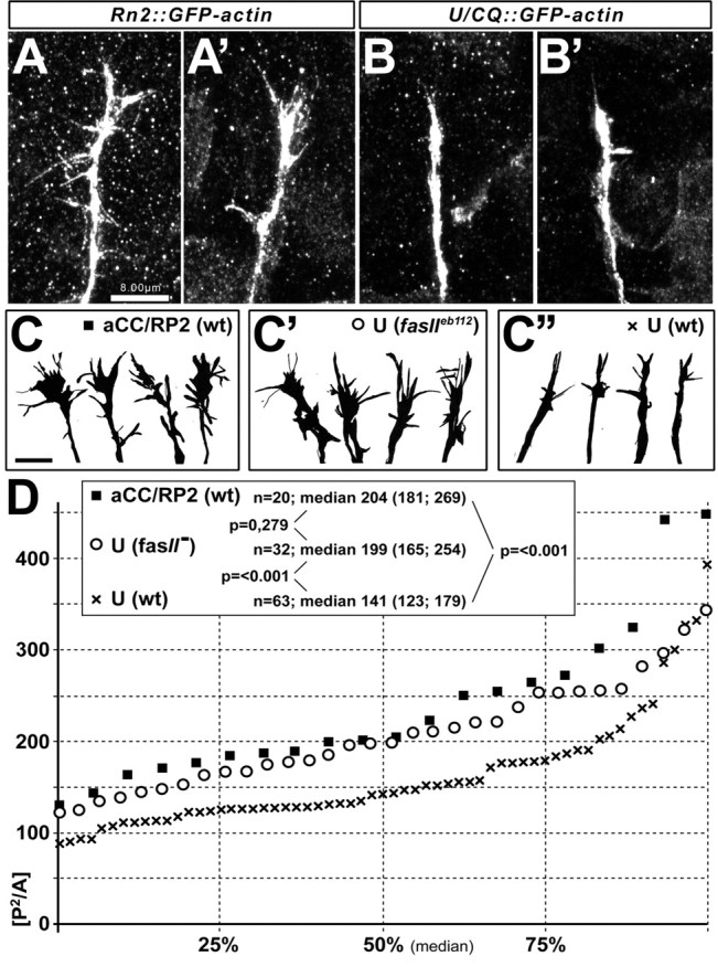
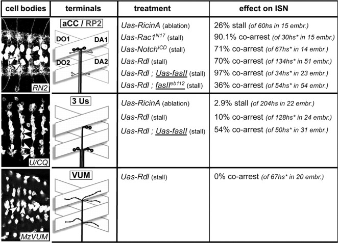
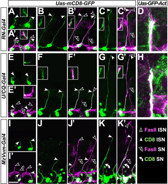
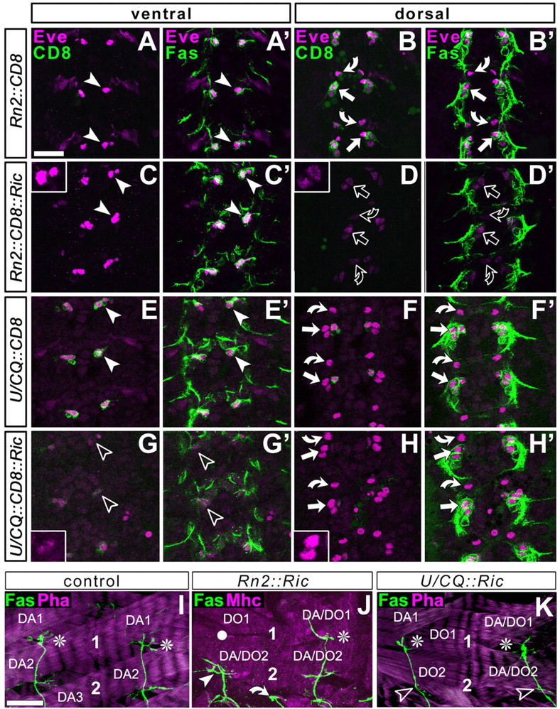
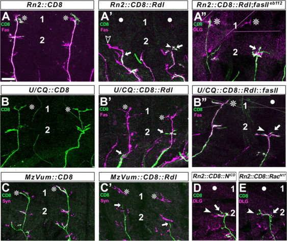
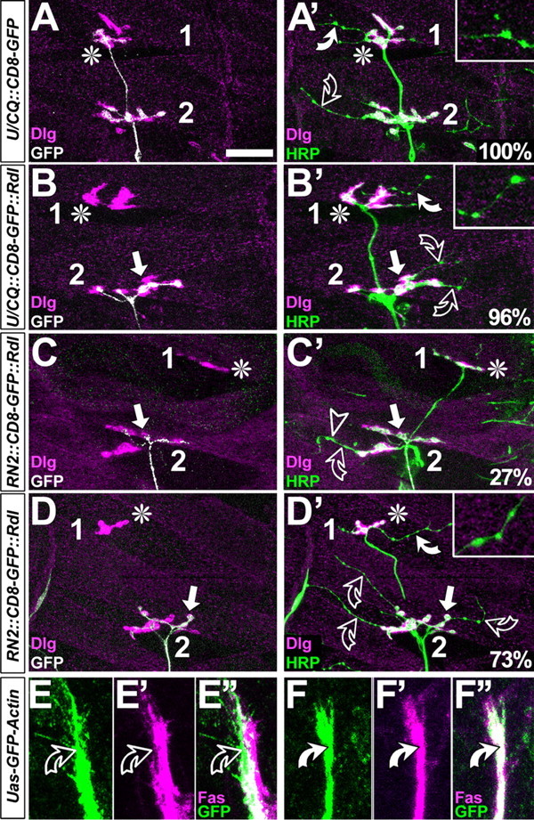
The phenomenon of pioneer neurons has been known for almost a century, but so far we have little insights into mechanisms and molecules involved. Here, we study the formation of the Drosophila intersegmental motor nerve (ISN). We show that aCC/RP2 and U motor neurons grow together at the leading front of the ISN. Nevertheless, aCC/RP2 neurons are the pioneers, and U neurons are the followers, because only aCC/RP2 neurons effectively influence growth of the ISN. We also show that this influence depends on the neural cell adhesion molecule homolog FasciclinII. First, ablation of aCC/RP2 has a stronger impact on ISN growth than U ablation. Second, strong growth-influencing capabilities of aCC/RP2 are revealed with a stalling approach we used: when aCC/RP2 motor axons are stalled specifically, the entire ISN (including the U neurons) coarrests, demonstrating that aCC/RP2 neurons influence the behavior of U growth cones. In contrast, stalled U neurons do not have the same influence on other ISN motor neurons. The influence on ISN growth requires FasciclinII: targeted expression of FasciclinII in U neurons increases their influence on the ISN, whereas a FasciclinII loss-of-function background reduces ISN coarrest with stalled aCC/RP2 axons. The qualitative differences of both neuron groups are confirmed through our findings that aCC/RP2 growth cones are wider and more complex than those of U neurons. However, U growth cones adopt aCC/RP2-like wider shapes in a FasciclinII loss-of-function background. Therefore, FasciclinII is to a degree required and sufficient for pioneer-follower interactions, but its mode of action cannot be explained merely through an equally bidirectional adhesive interaction.


Figures

Figure 6.
Figure 6.
Shapes of growth cones distinguish pioneers from followers and are influenced by FasciclinII. Whereas growth cones of aCC/RP2 neurons show numerous filopodia (A, A, C), growth cones of U neurons are narrow and poor in filopodia (B, B, C″; visualized with Rn2-Gal4- or U/CQ-Gal4-driven GFP-actin, respectively). Camera lucida drawings of growth cones (C-C″) were obtained from aCC/RP2 neurons in wild type (wt) (C, black square), U neurons in fasciclinIIeb112 loss-of-function mutant background (fasII) (C′, circle), or U neurons in wild-type embryos (C″, black cross). Their complexity index, P2/A, is plotted as a graph in D showing the median at 50% and interquartile ranges at 25 and 75%; the box in D contains sample numbers of measured growth cones (n), values of the respective medians (interquartile ranges in brackets separated by a semicolon), and significance values (p) obtained by Mann-Whitney rank sum tests (comparing the respective groups indicated by lines). Whereas U growth cones are significantly less complex than those of aCC/RP2 neurons in wild-type background, they are similarly complex in fasciclinIIeb112 mutant background. Scale bars, 8 μm.
Figure 1.
Figure 1.
Presentation of the used Gal4 driver lines and summary of the results obtained with them. Pictures in the left column (“cell bodies”) show ventral nerve cords of embryos at stage 17 (anterior left) expressing CD8-GFP targeted by RN2-Gal4, U/CQ-Gal4, or MzVum-Gal4, respectively; cell bodies of aCC/RP2, U, and VUM neurons are reliably stained (arrowheads) as are their terminals on most dorsal muscles. The second column from the left (“terminals”) illustrates the terminals of efferent neurons (named in box at the top) targeted by the respective driver lines and their dorsal target muscles [D, dorsal; A, acute; O, oblique; according to Bate (1993)]. The middle column (“treatment”) indicates the experiments performed with the respective Gal4 lines, i.e., targeted expression of “UAS-ricinA”, “UAS-Rdl”, “UAS-Rac1N17” or “UAS-NotchICD” (their effect on the targeted motor neurons is indicated in parentheses); UAS-Rdl was expressed in different backgrounds (underlined), i.e., “fasIIeb112” (fasciclinIIeb112 mutant background) and “UAS-fasII” (targeted coexpression of FasciclinII). The respective “effect on ISN” was analyzed in late stage 17 embryos and is given as a percentage of stall or coarrest of the ISN; numbers of analyzed hemisegments (hs) and embryos (embr.) are given in parentheses; hs* refers to only those hemisegments in which the targeted motor axons were stalled.
Figure 2.
Figure 2.
U and aCC/RP2 motor axons grow at the leading edge of the ISN. The ISN was analyzed at different time points (A, E, and I, stage 13; B, F, and J, stage 14; C, D, G, H, and K, stage 15). Preparations are labeled with anti-FasciclinII (FasII; magenta throughout); in addition, the different motor neurons (respective Gal4 lines indicated in boxes on the left) display targeted expression of mCD8-GFP or GFP-actin (green, as indicated at the top). Picture pairs show mCD8-GFP alone (no prime) or double-labeled with FasciclinII (prime). Nerve tips of the entire ISN are indicated by open arrowheads, tips of mCD8-labeled motor axons are indicated by white arrowheads, and positions of white arrowheads are shown as close-ups. Note that aCC/RP2 neurons grow at the leading edge of the ISN at all analyzed stages (A-C′), U neurons are delayed initially (E′) but grow at the leading edge later on (F,G′), and VUM neurons stay behind at all developmental stages (I-K′; 100% of cases; n = 45 from 10 embryos). Because mCD8-GFP often fails to show details such as filopodia, experiments were repeated with GFP-actin expression that likewise reveal that aCC/RP2 filopodia can be seen at the very tip of the FasciclinII-labeled ISN in 91% of cases (D; n = 22 hemisegments of 8 embryos); U axons grow at the ISN tip in 70% of cases (H; n = 45 hemisegments of 10 embryos). A second outgrowing nerve (bent open arrows) represents the substantia nigra pars compacta as suggested by the presence of MzVUM-Gal4 targeted axons (white curved arrow in K, K′) (Landgraf et al., 2003). Scale bar, 20 μm.
Figure 3.
Figure 3.
Ablation studies using targeted expression of ricinA. The CNS at embryonic stage 13 (A-H′) or dorsal muscle fields at late stage 17 (I-K) are labeled as indicated at the top left: CD8, Gal4-induced mCD8-GFP (green in first and third column); Eve, the transcription factor Even-skipped (magenta in A-H′); Fas, FasciclinII (green in second and fourth column and bottom row); Mhc, the muscle marker myosin heavy chain (magenta in J); Pha, actin labeled with phalloidin (magenta in I, K). A-H′, Views of the horizontal plane of an abdominal CNS (anterior up) at embryonic stage 13 (the early motor axonal growth phase) visualized at ventral (left two columns) or dorsal (right two columns) levels, respectively, as indicated in the boxes at the top. Cell bodies of ventral U neurons (arrowheads), dorsal aCCs (straight arrows), and dorsal RP2s (curved arrows) were visualized with three independent markers: FasciclinII and Even-skipped are expressed in all of these neurons (Grenningloh et al., 1991; Broadus et al., 1995); mCD8-GFP is expressed only in those neurons targeted by the respective Gal4 line (genetic constellation indicated on left side). RN2-Gal4 targets CD8 expression to dorsal aCC/pCC and RP2 neurons (B) but not to ventral U neurons (A); U/CQ-Gal4 mediated expression occurs in ventral U neurons (E) but not in aCC/pCC and RP2 neurons (F). If these Gal4 lines are used to coexpress ricinA (Ric) with CD8, expression of all three markers is severely affected, but only in the targeted neurons (open symbols inD, D′ and G, G′; see especially the granular and weak occurrence of Eve; insets); furthermore, the Fasciclin II-labeled motor nerve is much thinner, especially after ablation of the larger group of U neurons (data not shown). I-K, Dorsal muscle fields of two consecutive hemisegments, respectively, at the end of embryogenesis in control animals or specimens with neuron-specific expression of ricinA. 1 and 2 indicate motor neuronal terminals on DA1/DO1 and DA2/DO2 muscles, respectively (some muscles indicated for orientation; compare Fig. 1). In controls and many cases of ricinA-ablated neurons, the ISN (shown with FasciclinII, green) grows to full length (white asterisks); in 26% of cases in which a CC/RP2 are ablated, ISNs stall in dorsolateral areas (white arrowhead in J; white circles indicate noninnervated muscles), whereas only 2.9% of ISNs stall in the case of U ablation (K, open arrowheads indicate absence of U terminals on DA/DO2 muscles); curved arrow indicates transverse nerve in J. Scale bar: (in A) A-H′, 20 μm; (in I) I-K, 16 μm.
Figure 4.
Figure 4.
Axons of aCC/RP2 motor neurons can influence ISN formation. Figures show dorsal muscle fields of one or two consecutive hemisegments in late embryos (compare Fig. 1) (1 and 2 indicate position of terminals on DA1/DO1 and DA/DO2 muscles) with the terminals or axons of the entire ISN in magenta [visualized with FasciclinII (Fas), synapsin (Syn), or Discs large as indicated at the top left; see Results for details] and the Rn2-Gal4, U/CQ-Gal4, or MzVum-Gal4 targeted axons in green (visualized via CD8 expression). Normally, the CD8-labeled terminals of aCC/RP2 neurons (A), U neurons (B), and VUMs (C) reach most dorsal positions (asterisks). If Rdl is expressed in these neurons (A-C′), their axons stall frequently (straight arrows); entire ISNs tend to coarrest with stalled aCC/RP2s (A′; white circles indicate noninnervated muscles; arrowheads point at tips of ISNs, open arrowhead in A′ indicates a case in which axons escaped aCC/RP2 but stalled thereafter, as in ablation experiments). ISNs mostly escape from stalled U neurons and VUMs (B, C′, asterisks; ISN coarrestin Rn2::Rdl = 70%; U/CQ::Rdl = 10%; MzVum::Rdl = 0%). Coarrest of ISNs with stalled aCC/RP2 motor axons is severely suppressed in fasciclinIIeb112 loss-of-function mutant background as shown for the right segment in A″ where the aCC/RP2 axons (green) are stalled, whereas Dlg-labeled terminals on dorsal muscles (magenta) are visible (ISN coarrest in Rn2::Rdl; fasIIeb112 = 36%). In contrast, ISN coarrest with stalled U neurons is severely increased, if FasciclinII is coexpressed with Rdl (right axon in B″; ISN coarrest in U/CQ::Rdl::fasII = 54%). Targeted expression of activated Notch (NICD; D) or dominant-negative DRac1 (RacN17; E) in aCC/RP2s likewise leads to axon stall and coarrest of the ISN, confirming the findings with Rdl (see Fig. 1 for details). Scale bar, 16 μm.
Figure 5.
Figure 5.
FasciclinII might not act merely as a homophilic adhesion factor. A-D′, Dorsal muscle fields of embryos at the late first and early second instar larval stage; symbols are as in Figure 4 and stainings are indicated at the bottom left. In control hemisegments, CD8-GFP-labeled U neurons grow to the most dorsal muscle (asterisk in A), as does the entire nerve (HRP in A′). Whereas motor neuronal type 1 terminals display large boutons and are double-labeled with Dlg and HRP (white color in A′), terminals of type 2 modulatory VUM neurons are slim and lack Dlg [throughout this figure, white curved arrows and closeups show type 2 terminals on DA1/DO1 muscles; open curved arrows show type 2 terminals on DA2/DO2; for terminal classification, compare Landgraf et al. (2003)]. If U neurons are stalled (arrow in B, B′), terminals of remaining ISN type 1 terminals are almost always present (asterisk in B), and VUM neurons are seen in most dorsal positions in 96% of these cases. If aCC/RP2 neurons are stalled (white arrows in C-D′), nearly all ISN neurons coarrest (Fig 4 A,D,E), but in 30% of cases U neurons escape (asterisks in C-D′); in these cases, VUM neurons are absent on dorsal muscles in 27% (C′) and present in the remaining 73% (D′). E-F″, Growth cones and axons of aCC/RP2 (green in C-C″; RN2::GFP-actin) mostly show low levels of FasciclinII (magenta) on exposed surfaces (curved open arrows), whereas GFP-Actin expressed in U neurons (green in D-D″; U/CQ::GFP-actin) is always congruent with high levels of FasciclinII (curved white arrows). Scale bar: A-B′, 25 μm; C-D″, 8 μm.

References

    1. Bak M, Fraser SE (2003) Axon fasciculation and differences in midline kinetics between pioneer and follower axons within commissural fascicles. Development 130: 4999-5008. - PubMed
    1. Bate M (1993) The mesoderm and its derivatives. In: The development of Drosophila melanogaster (Bate M, Martínez Arias A, eds), pp 1013-1090. Cold Spring Harbor, NY: Cold Spring Harbor Laboratory.
    1. Boschert U, Ramos RG, Tix S, Technau GM, Fischbach KF (1990) Genetic and developmental analysis of irreC, a genetic function required for optic chiasm formation in Drosophila. J Neurogenet 6: 153-171. - PubMed
    1. Bowie JE, Young IT (1977) An analysis technique for biological shape-II. Acta Cytol 21: 455-464. - PubMed
    1. Brand A, Perrimon N (1993) Targeted gene expression as a means of altering cell fates and generating dominant phenotypes. Development 118: 401-415. - PubMed

Publication types

LinkOut - more resources