. 2005 Jan 5;25(1):118-29.
doi: 10.1523/JNEUROSCI.3571-04.2005.

The rod photoreceptor-specific nuclear receptor Nr2e3 represses transcription of multiple cone-specific genes

Affiliations

The rod photoreceptor-specific nuclear receptor Nr2e3 represses transcription of multiple cone-specific genes

Jichao Chen et al. J Neurosci. .

Abstract

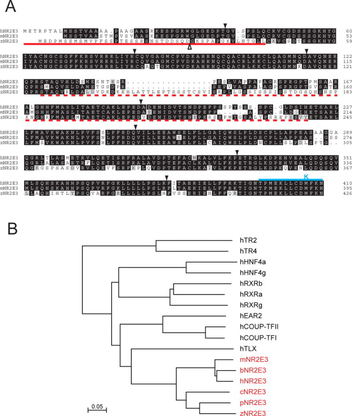
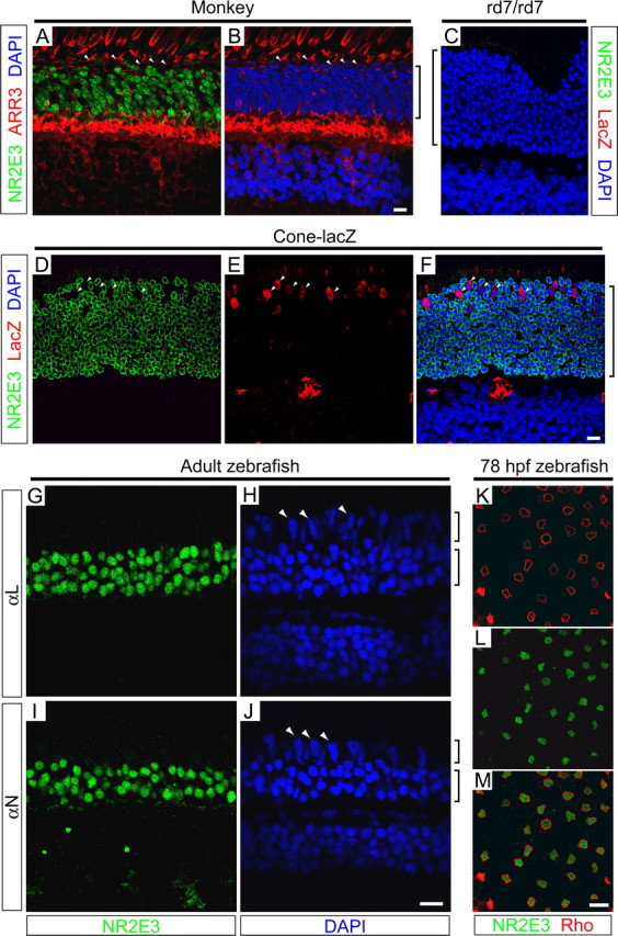
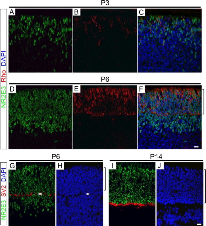
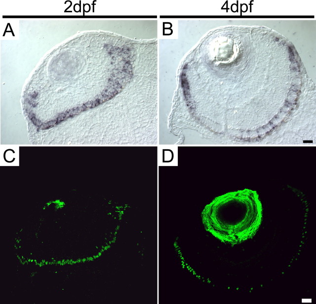
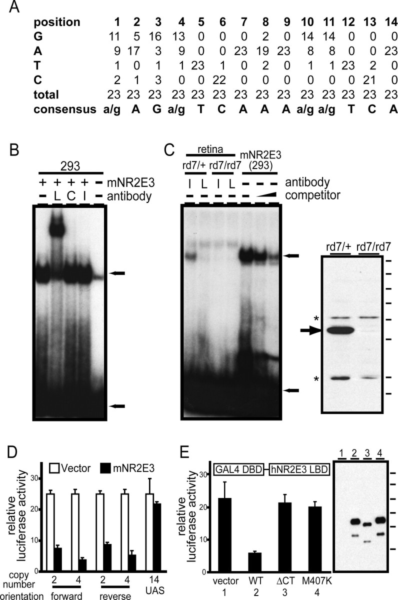
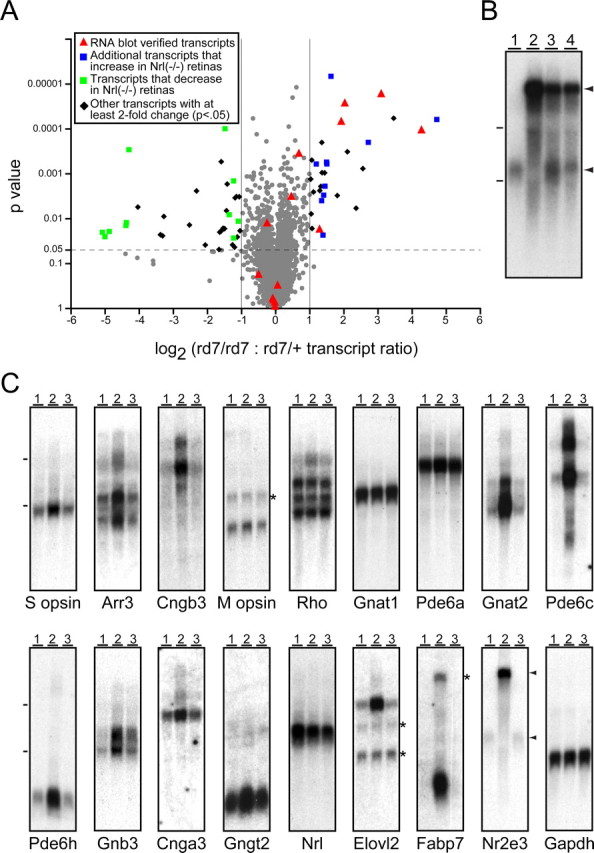
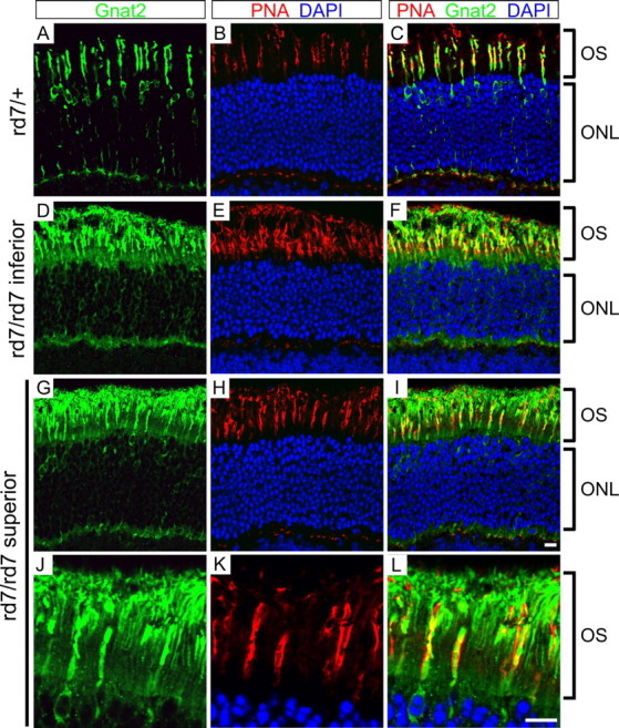
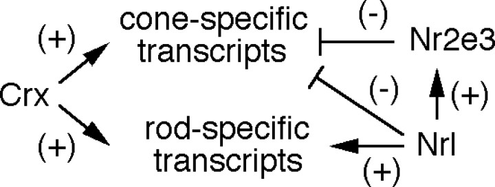
This study addresses one genetic regulatory mechanism that establishes the distinct identities of rod and cone photoreceptors. Previous work has shown that mutations in either humans or mice in the gene coding for photoreceptor-specific nuclear receptor Nr2e3 cause a progressive retinal degeneration characterized by increased numbers of short-wave cones. In the present work, we have examined the cellular and developmental pattern of Nr2e3 protein localization in mammals and fish, identified an optimal Nr2e3 DNA-binding site using cycles of binding to recombinant Nr2e3, characterized the transcriptional activity of wild type and one of the disease-associated point mutations in Nr2e3 in transfected cells, and characterized the transcriptional defects in the naturally occurring Nr2e3 mutant (rd7) mouse. These experiments indicate that in the mature vertebrate retina Nr2e3 is expressed exclusively in rods, that expression of Nr2e3 is one of the earliest events in the pathway of rod-specific photoreceptor development, and that Nr2e3 functions, either directly or indirectly, as a repressor of cone-specific genes in rod photoreceptor cells.


Figures

Figure 1.
Figure 1.
Comparison of Nr2e3 amino acid sequences among diverse vertebrates. A, Alignment of human, mouse, and zebrafish Nr2e3 sequences. Intron positions are shown with downward filled arrowheads. Except for the first intron in the zebrafish gene, shown by the upward open arrowhead, the intron locations are conserved. The solid red underline indicates immunogens used to generate anti-mouse and -zebrafish Nr2e3 N-terminal antibodies. The dashed red underline indicates immunogens used to generate anti-mouse and anti-zebrafish Nr2e3 linker region antibodies and the region included in the fusion protein used to affinity purify anti-human Nr2e3 linker region antibodies. The solid blue line and K above the C-terminal region indicate C-terminal and M407K truncation mutants, respectively, studied in human Nr2e3. B, Dendrogram showing the percent amino acid identities among Nr2e3 orthologs from various vertebrates and among the most closely related nuclear receptors from humans. Scale bar, 5% divergence. b, Bovine; c, chicken; h, human; m, mouse; p, pufferfish (Fugu rubripes); z, zebrafish.
Figure 2.
Figure 2.
Immunolocalization of Nr2e3 to rod photoreceptors in monkey, mouse, and zebrafish retinas. A, B, Macaque retina immunostained for Nr2e3 and cone arrestin (ARR3; mAb 7G6) and counterstained with 4′, 6-diamidino-2-phenylindole (DAPI). C-F, rd7/rd7 (C) and transgenic cone-β galactosidase (D-F) mouse retinas immunostained for Nr2e3 and β-galactosidase and counterstained with DAPI. The specificities of mNr2e3 and β-galactosidase antibodies are demonstrated by the absence of immunostaining in the nontransgenic rd7/rd7 retina. The large red object in the outer plexiform layer in E and F is a blood vessel in which intravascular mouse IgG is stained with the Alexa 594-conjugated anti-mouse secondary antibody. G-J, Zebrafish retinas immunostained with antibodies to amino acids 1-45 (αN) and 125-239 (αL) of zNr2e3 and counterstained with DAPI. K-M, Flat mount of zebrafish retina at 78 hpf immunostained for rhodopsin and zNr2e3 shows that, in the central retina at this time point, Nr2e3 is observed exclusively in rods. In each of the species examined, Nr2e3 immunostaining is confined to rod nuclei; cone nuclei (A, B, D-F, H, J, white arrowheads) do not contain Nr2e3. Brackets adjacent to B, C, and F indicate the outer nuclear layer; brackets adjacent to H and J indicate the cone (top bracket) and rod (bottom bracket) nuclear layers. Scale bars, 10 μm.
Figure 3.
Figure 3.
Nr2e3 accumulates in rod precursors in the developing mouse retina. A-F, P3 (A-C) and P6 (D-F) mouse retinas immunostained for Nr2e3 and rhodopsin and counterstained with DAPI. G-J, P6 (G, H) and P14 (I, J) mouse retinas immunostained for Nr2e3 and SV2 and counterstained with DAPI. Only the outer half of each retina, containing the future inner and outer nuclear layers, is shown. Brackets adjacent to F, H, and J indicate the developing outer nuclear layer. Arrowheads in G and H indicate the developing outer plexiform layer. All rhodopsin-containing cells also contain Nr2e3, but additional cells, presumably early rod precursors, contain Nr2e3 but do not contain rhodopsin. Scale bars, 10 μm.
Figure 4.
Figure 4.
Transient Nr2e3 expression in cones in the developing zebrafish retina. In situ hybridization (A, B) and immunostaining (C, D) for Nr2e3 at 2 and 4 dpf is shown. Immunostaining of the lens is nonspecific. Cells throughout the photoreceptor layer are stained at 2 dpf, but only rods express Nr2e3 in the most mature (central) region of the retina at 4 dpf. Scale bars, 20 μm.
Figure 5.
Figure 5.
An optimal Nr2e3-binding site mediates Nr2e3-dependent transcriptional repression in transfected cells. A, Compilation of 23 cloned oligonucleotide sequences after three rounds of selection with a GST fusion to full-length mNr2e3. B, EMSA with mNr2e3 produced in transfected 293 cells using the DNA duplex probe defined by Kobayashi et al. (1999) (see Materials and Methods). Antibodies directed against the hNr2e3 linker region (L) produce a supershift; antibodies directed against the C-terminal 73 amino acids of hNr2e3 (C) or against an irrelevant protein (photoreceptor cadherin; I) do not produce a mobility shift. Extracts from untransfected 293 cells (-) produce a weak EMSA band that has a slightly higher electrophoretic mobility than that produced by mNr2e3. C, Left panel, EMSA using an optimal DNA duplex probe determined by in vitro selection (see Materials and Methods). Left four lanes, EMSA using retina extracts from rd7/+ or rd7/rd7 littermates incubated with anti-hNr2e3 linker region or irrelevant antibodies. Right three lanes, EMSA with mNr2e3 produced in transfected 293 cells in the presence of increasing quantities (0.5 and 2.5 μm) of the same optimal DNA duplex as competitor. Top and bottom arrows, Bound and free probe, respectively. Right panel, Specificity of the anti-hNr2e3 linker region antibodies used for super shifting experiments determined by immunoblotting of retina proteins from rd7/+ or rd7/rd7 littermates. Arrow, Nr2e3; asterisks, cross-reacting proteins. Molecular mass standards (from top to bottom): 183, 114, 81, 64, 50, 37, 26, and 20 kDa. D, Cotransfection of 293 cells with a vector (pRK5) directing the expression of full-length mNr2e3 or with pRK5 alone, together with a luciferase reporter under the control of a minimal herpes simplex virus thymidine kinase promoter 3′ of the indicated number and orientation of optimal Nr2e3-binding sites determined by in vitro selection. A control reporter carries 14 Gal4 UAS sites in place of the Nr2e3 sites. Nr2e3 represses expression in a target-site-dependent manner. E, Cotransfection of 293 cells with pRK5 directing the expression of a Gal4DBD-hNr2e3LBD fusion protein together with a luciferase reporter under the control of a minimal herpes simplex virus thymidine kinase promoter 3′ of 14 tandem Gal4 UAS sites. The hNr2e3 LBD corresponds to amino acids 126-410. Repression of the reporter is seen with the WT Nr2e3 LBD sequence but not with a C-terminal truncation of 13 amino acids (ΔCT), substitution mutation M407K, or empty pRK5 vector. Right panel, Immunoblot of the different Gal4-Nr2e3LBD constructs expressed in 293 cells with anti-hNr2e3 antibodies. Lanes 1-4 refer to the four numbered samples in the graph. Each fusion protein shows similar levels of cleavage between the Gal4 and Nr2e3 domains, producing a lower-molecular weight Nr2e3LBD polypeptide. Molecular mass standards (from top to bottom): 183, 114, 81, 64, 50, and 37 kDa. Error bars in D and E indicate SDs of at least three experiments.
Figure 6.
Figure 6.
In situ hybridization reveals distinct patterns of cone-specific gene expression in rd7/rd7 retinas. A-L, In situ hybridization to retinas obtained from 6-week-old rd7/+ (left) or rd7/rd7 (right) littermates with probes for the indicated rod and cone transcripts. A-D, Rod transcripts with no detectable alteration in expression in the rd7/rd7 retina. E-H, Cone transcripts that are mostly excluded from rods in both rd7/ + and rd7/rd7 retinas. I-L, Conetranscripts that are expressed in rods in the rd7/rd7 retina. The sections with S-opsin (E) and M-opsin (H) hybridizations are derived from inferior and superior retina, respectively, the regions with the highest concentrations of S- and M-cones. M, N, Hybridization to 6-week-old +/+ and rd7/rd7 retinas with an Nr2e3 probe. O, P, Superior region of rd7/rd7 retinas at the indicated ages hybridized with S-opsin (left) or M-opsin (right) probes. As in the WT retina, S-cones are present at low density in the superior half of the rd7/rd7 retina, but in the rd7/rd7 retina, they reside in both normal and abnormal (i.e., inner) regions of the outer nuclear layer (ONL). In the rd7/rd7 retina, M-cone cell bodies are initially found through out the ONL, and this distribution progressively shifts to the outer edge of the ONL, as seen by comparing M-opsin hybridization patterns at P14 versus 6 weeks (P, H). Each panel shows the full thickness of the retina, with the retinal pigment epithelium at the top and the ganglion cell layer at the bottom. The vertical brackets indicate the extent of the ONL. Characteristic retinal folds are seen in the rd7/rd7 sections, but the relative composition of photoreceptor cell types does not appear to differ between folded and normal regions of the retina. For each probe, the pair of sections of different genotypes was processed on the same slide; therefore, relative signal intensities directly reflect differences in transcript abundance. Chromogenic substrate development times differed among probes. Scale bar, 20 μm.
Figure 7.
Figure 7.
Microarray and RNA blot hybridization shows selectivity in rd7/rd7 misregulation of cone-specific transcripts. A, Scatterplot showing the rd7/rd7:rd7/ + abundance ratio for P30 retina transcripts averaged over three independent microarray experiments using the U74Av2 chip from Affymetrix. Abundance ratios are plotted on the x-axis and are stratified by p value on the y-axis. Most transcripts change by less than twofold and are represented by gray circles. Transcripts tested by RNA blotting (C) are represented by red triangles. Additional upregulated transcripts that are also upregulated in Nrl(-/-) retinas (Yoshida et al., 2004) are shown as blue squares, and down regulated transcripts that are also downregulated in Nrl(-/-) retinas (Yoshida et al., 2004) are shown as green squares. Other transcripts with at least a twofold change and a p < 0.05 are shown as black diamonds. We note that several photoreceptor transcripts (e.g., Nr2e3, Cngb3, Pde6c, Pde6h, Arr3) are not represented on the U74Av2 chip. B, Equal quantities of total RNA from 1- to 2-month-old WT (lane 1), rd7/rd7 (lane 2), rd7/+ (lane 3), and a 1:1 mixture of WT and rd7/rd7 (lane 4) retinas hybridized with an Nr2e3 probe. Arrowheads to the right of the Nr2e3 blots in B and C indicate the normal Nr2e3 transcript (bottom arrowhead) and the high-molecular weight variant Nr2e3 transcript (top arrowhead). C, Total RNA from 1- to 2-month-old WT (lane 1), rd7/rd7 (lane 2), and prCAD(-/-) (lane 3) retinas hybridized with the indicated probes. At these ages, the rd7/rd7 and prCAD(-/-) retinas show only a minimal loss of photoreceptors. Dashes to the left of the blots in B and C show the mobilities of the large and small ribosomal RNAs. The asterisks to the right of several blots show the locations of residual hybridization signals from earlier experiments. Exposure times are not matched across blots.
Figure 8.
Figure 8.
The α subunit of cone transducin localizes to both cone and rod photoreceptors in rd7/rd7 retinas. A-C, rd7/+ retinas immunostained for the α subunit of cone transducin (Gnat2; green) and costained with PNA (red) and DAPI (blue). D-L, rd7/rd7 stained as in A-C. D-F, The inferior rd7/rd7 retina has an approximately twofold higher density of S-cones than an rd7/+ retina. G-L, the superior rd7/rd7 retina has a normal concentration of M-cones. The outer segment region of G-I is enlarged in J-L, showing the localization of cone transducin α to both rod and cone outer segments (OS). Retinas were harvested from littermates at 6 weeks of age. PNA marks each cone sheath, an extracellular matrix structure that encompasses the cone OS and extends distal to it. Scale bar, 10 μm.
Figure 9.
Figure 9.
Model for transcriptional control in rod photoreceptors. Crx activates the transcription of both rod- and cone-specific genes (Furukawa et al., 1999), and Nrl activates the transcription of rod-specific genes, including Nr2e3 (Mears et al., 2001). Both Nrl (Mears et al., 2001) and Nr2e3 repress the transcription of cone-specific genes. In the mouse retina, Crx transcripts appear at ∼E12.5 (Furukawa et al., 1997), Nrl transcripts appear at ∼E14.5 (Liu et al., 1996), and Nr2e3 transcripts appear at ∼E16.5 (Cheng et al., 2004), suggesting a temporal hierarchy with Crx and Nrl above Nr2e3.

References

    1. Akhmedov NB, Piriev NI, Chang B, Rapoport AL, Hawes NL, Nishina PM, Nusinowitz S, Heckenlively JR, Roderick TH, Kozak CA, Danciger M, Davisson MT, Farber DB (2000) A deletion in a photoreceptor-specific nuclear receptor mRNA causes retinal degeneration in the rd7 mouse. Proc Natl Acad Sci USA 97: 5551-5556. - PMC - PubMed
    1. Anderson RE, Maude MB, Bok D (2001) Low docosahexaenoic acid levels in rod outer segment membranes of mice with rds/peripherin and P216L peripherin mutations. Invest Ophthalmol Vis Sci 42: 1715-1720. - PubMed
    1. Arannda A, Pascual A (2001) Nuclear hormone receptors and gene expression. Physiol Rev 81: 1269-1304. - PubMed
    1. Bessant DA, Payne AM, Mitton KP, Wang QL, Swain PK, Plant C, Bird AC, Zack DJ, Swaroop A, Bhattacharya SS (1999) A mutation in NRL is associated with autosomal dominant retinitis pigmentosa. Nat Genet 21: 355-356. - PubMed
    1. Bicknell IR, Darrow R, Barsalou L, Fliesler SJ, Organisciak DT (2002) Alterations in retinal rod outer segment fatty acids and light-damage susceptibility in P23H rats. Mol Vis 8: 333-340. - PubMed

Publication types

MeSH terms