

Comparative Study
doi: 10.1523/JNEUROSCI.3156-04.2005.
Pup suckling is more rewarding than cocaine: evidence from functional magnetic resonance imaging and three-dimensional computational analysis
Affiliations
- PMID: 15634776
- PMCID: PMC6725197
- DOI: 10.1523/JNEUROSCI.3156-04.2005
Item in Clipboard
Comparative Study
Pup suckling is more rewarding than cocaine: evidence from functional magnetic resonance imaging and three-dimensional computational analysis
J Neurosci.
.
Abstract
Nursing has reciprocal benefits for both mother and infant, helping to promote maternal behavior and bonding. To test the "rewarding" nature of nursing, functional magnetic resonance imaging was used to map brain activity in lactating dams exposed to their suckling pups versus cocaine. Suckling stimulation in lactating dams and cocaine exposure in virgin females activated the dopamine reward system. In contrast, lactating dams exposed to cocaine instead of pups showed a suppression of brain activity in the reward system. These data support the notion that pup stimulation is more reinforcing than cocaine, underscoring the importance of pup seeking over other rewarding stimuli during lactation.
Figures
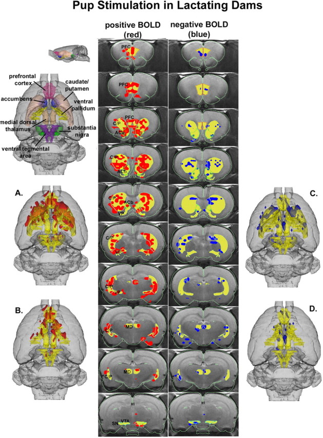
3D activational map showing pup stimulation in lactating dams. Top left picture is a translucent shell of the brain viewed from a dorsal perspective showing color-coded VOIs corresponding to anatomical geometries of the mesocorticolimbic and nigrostriatal dopamine systems. For orientation, a smaller rendering of the same picture is shown from a lateral perspective. Colors have been melded into a single functional VOI (yellow) showing localization of positive (red) and negative (blue) BOLD signal changes with pup stimulation. A includes both dopamine systems, whereas B has masked the caudate-putamen (C/P) and substantia nigra (SN) comprising the nigrostriatal dopamine system, revealing the mesocorticolimbic or reward system. C and D are the corresponding negative BOLD images for each functional volume, respectively. The middle columns show traditional activation maps of contiguous brain sections with labeled regions of interest. The significantly activated voxels are the composite of all six subjects overlaid on one subject used as an anatomical template. The location of every voxel in any subject whose p value was <0.05 after false-positive filtering for ± BOLD signal change is included in the composite analysis and located within the region of interest. PFC, Prefrontal cortex; ACb, accumbens; VP, ventral pallidum; MD, medial dorsal thalamus.
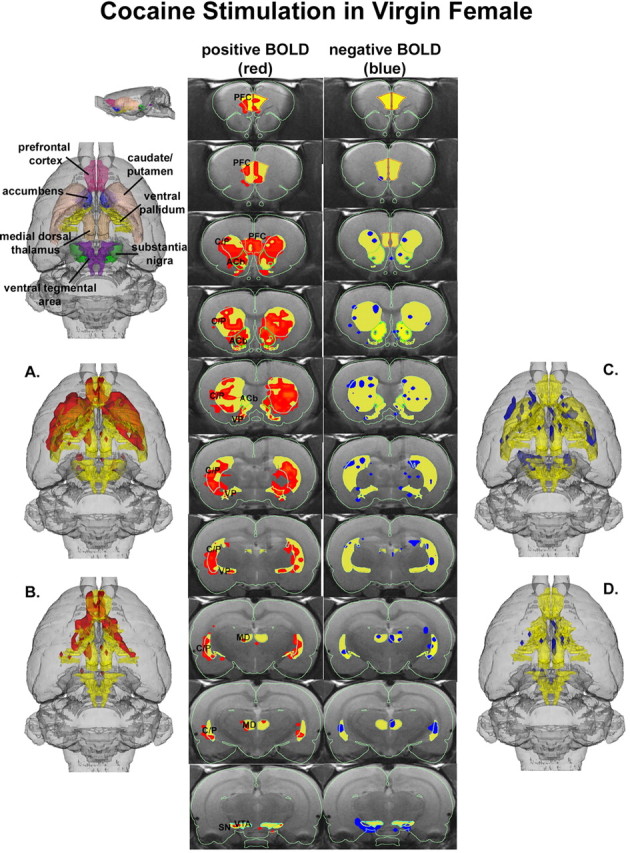
3D activational map showing cocaine stimulation in virgin females. Same as Figure 1. C/P, Caudate-putamen; SN, substantia nigra; PFC, prefrontal cortex; ACb, accumbens; VP, ventral pallidum; MD, medial dorsal thalamus.
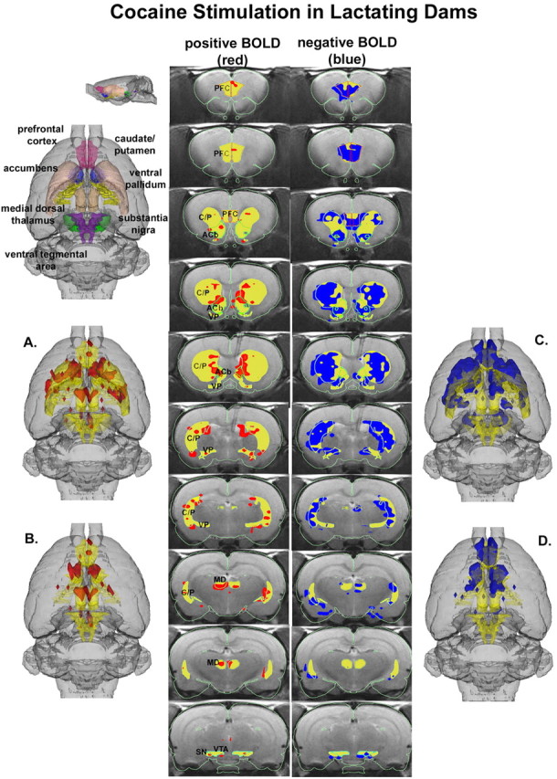
3D activational map showing cocaine stimulation in lactating dams. Same as Figure 1. C/P, Caudate-putamen; SN, substantia nigra; PFC, prefrontal cortex; ACb, accumbens; VP, ventral pallidum; MD, medial dorsal thalamus.
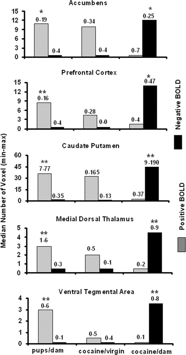
Volumes of activation. The bar graphs show the median number of voxels with a significant change in BOLD signal intensity with a p value <0.05 after applying the false-positive filter for the volumes of interest noted. Numbers above each bar represent the minimum and maximum from six animals. There was no significant differences in the number of positive (gray) or negative (black) voxels between pups- dam and cocaine-virgin conditions. *p < 0.05; **p < 0.01, using a Bonferroni correction for a three-group comparison.
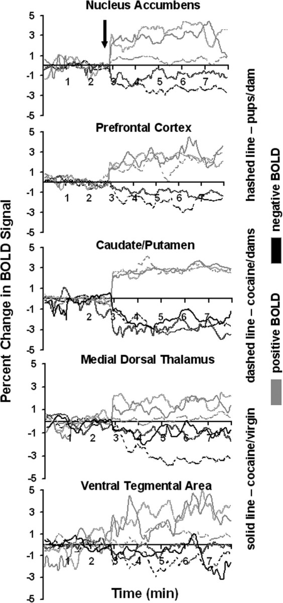
BOLD signal changes over time. The time course plots show the percentage change in positive (gray) and negative (black) BOLD signal after stimulus presentation (arrow) for the corresponding volumes of interest. The stimuli were either exposure to pups for the duration of 5 min or a bolus ICV injection of cocaine (20 μg/20 μl) followed by 5 min of data acquisition.
References
-
- Fleming AS, Korsmit M, Deller M (1994) Rat pups are potent reinforcers to the maternal animal: effects of experience, parity, hormones and dopamine function. Psychobiology (Austin, Tex) 22: 44-53.
-
- Genovese CR, Lazar NA, Nichols T (2002) Thresholding of statistical maps in functional neuroimaging using the false discovery rate. NeuroImage 15: 870-878. - PubMed
Publication types
MeSH terms
Substances
Grants and funding
LinkOut - more resources
Full Text Sources