. 2005 Jan 5;25(1):184-91.
doi: 10.1523/JNEUROSCI.4269-04.2005.

Uncoupling protein-2 is critical for nigral dopamine cell survival in a mouse model of Parkinson's disease

Affiliations

Uncoupling protein-2 is critical for nigral dopamine cell survival in a mouse model of Parkinson's disease

Zane B Andrews et al. J Neurosci. .

Erratum in

  • J Neurosci. 2005 Feb 23;25(8):table of contents

Abstract

Mitochondrial uncoupling proteins dissociate ATP synthesis from oxygen consumption in mitochondria and suppress free-radical production. We show that genetic manipulation of uncoupling protein-2 (UCP2) directly affects substantia nigra dopamine cell function. Overexpression of UCP2 increases mitochondrial uncoupling, whereas deletion of UCP2 reduces uncoupling in the substantia nigra-ventral tegmental area. Overexpression of UCP2 decreased reactive oxygen species (ROS) production, which was measured using dihydroethidium because it is specifically oxidized to fluorescent ethidium by the superoxide anion, whereas mice lacking UCP2 exhibited increased ROS relative to wild-type controls. Unbiased electron microscopic analysis revealed that the elevation of in situ mitochondrial ROS production in UCP2 knock-out mice was inversely correlated with mitochondria number in dopamine neurons. Lack of UCP2 increased the sensitivity of dopamine neurons to 1-methyl-4-phenyl-1,2,5,6 tetrahydropyridine (MPTP), whereas UCP2 overexpression decreased MPTP-induced nigral dopamine cell loss. The present results expose the critical importance of UCP2 in normal nigral dopamine cell metabolism and offer a novel therapeutic target, UCP2, for the prevention/treatment of Parkinson's disease.


Figures
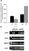
Figure 1.
Figure 1.
Fatty acid-induced uncoupling in UCP2 WT/TG and WT/KO mice and UCP expressions. A, Exposure to the free fatty acid palmitate induces increased mitochondrial uncoupling in UCP2 TG mice when compared with WT controls. Asterisk indicates p < 0.001; n = 4. UCP2 KO mice show reduced mitochondrial uncoupling compared with WT controls. Asterisk indicates p = 0.002; n = 4. Data are expressed as the percentage of increase above oligomycin-induced state 4 respiration. Error bars represent SEM. B, Real-time PCR analysis of the SN revealed expressions of both UCP4 and BMCP1 mRNAs without significant differences between their expression levels in UCP2 KO, hUCP2-expressing mice an their respective wild-type littermates.
Figure 2.
Figure 2.
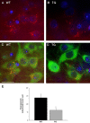
Free-radical production in TH neurons of the SNc in UCP2 WT/TG mice. A, B, Representative photomicrographs of in situ ROS production assessed by the oxidation of DHE to ethidium by formula image. Left photomicrographs show a representation of double exposure (A) for ethidium (red) and Hoechst nuclear staining (blue) or triple exposure (B) for ethidium, Hoechst nuclear staining, and TH immunofluorescence (green) in UCP2 WT mice. C, D, Representative findings of ethidium and Hoechst nuclear staining (C) with TH immunofluorescence (D) in UCP2 TG mice. Scale bar: (in C) A-D, 10 μm. E, Quantification of the number of red fluorescent mitochondria within TH cells revealed a significant decrease in UCP2-overexpressing animals compared with their WT controls (n = 4, n = 3, respectively). *Significant with respect to WT controls (p < 0.05). Error bars represent SEM.
Figure 3.
Figure 3.
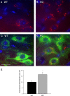
Free-radical production in TH neurons of the SNc in UCP2 WT/KO mice. A, B, Representative photomicrographs of in situ ROS production assessed by the oxidation of DHE to ethidium by formula image. Left photomicrographs show a representation of double exposure (A) for ethidium (red) and Hoechst nuclear staining (blue) or triple exposure (B) for ethidium, Hoechst nuclear staining, and TH immunofluorescence (green) in UCP2 WT mice. C, D, In UCP2 KO mice (right photomicrographs), representative findings for ethidium and Hoechst nuclear staining (C) with TH immunofluorescence (D). Scale bar: (in C) A-D, 10 μm. E, Quantification of the number of red fluorescent mitochondria within TH cells revealed a significant increase in UCP2 KO mice compared with their WT controls (n = 6, n = 5, respectively). *Significant with respect to WT controls (p < 0.05). Error bars represent SEM.
Figure 4.
Figure 4.
UCP2 KO mice have reduced mitochondria in SNc TH cells. A, Representative electron micrograph shows numerous mitochondria in UCP2 WT mice in the cytoplasm of immunoreactive TH cells examined with electron microscopy. B, Representative electron micrographs of UCP2 KO mice illustrate reduced mitochondrial number in the cytoplasm of immunoreactive TH cells examined with electron microscopy. Asterisks in A and B indicate mitochondria. Scale bar: (in B) A, B, 1 μm. C, Quantification of the number of mitochondria in UCP2 WT/TG and UCP2 WT/KO mice. Data are normalized to cytoplasmic area and expressed as mitochondrial number per square micrometer. UCP2 KO mice (n = 14) had reduced mitochondria within TH neurons compared with WT controls (n = 27; 0.460 ± 0.042 vs 0.318 ± 0.028; p < 0.05). There was no difference between UCP2 TG mice (n = 18) and their WT controls (n = 15; 0.448 ± 0.052 vs 0.440 ± 0.029, respectively). *Significant differences in mitochondria counts between UCP2 KO and wild-type animals. Error bars represent SEM.
Figure 5.
Figure 5.
MPTP-induced TH cell loss in UCP2 TG and WT mice. A-D, Dopamine cell number assessed by tyrosine hydroxylase immunocytochemistry showed decreased number of cells in MPTP-treated WT and UCP2 TG mice (n = 5, 6). Scale bar: (in B) A-D, 50 μm. E, Stereological quantification of dopamine cell number revealed that UCP2 TG mice had significantly (p < 0.05) more surviving neurons than their WT littermates after MPTP treatment. *Significant with respect to wild-type controls. F, Analysis of striatal dopamine levels showed equally diminished dopamine levels after MPTP injections in transgenic and wild-type animals. Error bars represent SEM.
Figure 6.
Figure 6.
MPTP-induced dopamine cell loss in UCP2 KO and WT mice. A-D, Dopamine cell number assessed by tyrosine hydroxylase immunocytochemistry showed decreased number of cells in MPTP-treated WT and UCP2 KO mice (n = 5, 6). Scale bar: (in B) A-D, 50 μm. E, Stereological quantification revealed that UCP2 KO mice had significantly (p < 0.05) less surviving dopamine neurons than their WT littermates after MPTP treatment. *Significant with respect to WT controls. F, Analysis of striatal dopamine levels showed diminished dopamine levels after MPTP injections in both wild-type and UCP2 KO animals. *Significant differences in striatal dopamine levels between UCP2 KO and wild-type animals. However, in MPTP-treated UCP2 KO animals, there was significantly (p < 0.05) lower levels of dopamine remaining in the striatum compared with the values of wild-type animals. Error bars represent SEM.

References

    1. Alleva R, Tomasetti M, Andera L, Gellert N, Borghi B, Weber C, Murphy MP, Neuzil J (2001) Coenzyme Q blocks biochemical but not receptor-mediated apoptosis by increasing mitochondrial antioxidant protection. FEBS Lett 503: 46-50. - PubMed
    1. Arsenijevic D, Onuma H, Pecqueur C, Raimbault S, Manning BS, Miroux B, Couplan E, Alves-Guerra MC, Goubern M, Surwit R, Bouillaud F, Richard D, Collins S, Ricquier D (2000) Disruption of the uncoupling protein-2 gene in mice reveals a role in immunity and reactive oxygen species production. Nat Genet 26: 435-439. - PubMed
    1. Beal MF, Matthews RT, Tieleman A, Shults CW (1998) Coenzyme Q10 attenuates the 1-methyl-4-phenyl-1,2,3, tetrahydropyridine (MPTP) induced loss of striatal dopamine and dopaminergic axons in aged mice. Brain Res 783: 109-114. - PubMed
    1. Bechmann I, Diano S, Warden CH, Bartfai T, Nitsch R, Horvath TL (2002) Brain mitochondrial uncoupling protein 2 (UCP2): a protective stress signal in neuronal injury. Biochem Pharmacol 64: 363-367. - PubMed
    1. Bindokas VP, Jordan J, Lee CC, Miller RJ (1996) Superoxide production in rat hippocampal neurons: selective imaging with hydroethidine. J Neurosci 16: 1324-1336. - PMC - PubMed

Publication types

MeSH terms