. 2005 Jan 5;25(1):252-9.
doi: 10.1523/JNEUROSCI.3054-04.2005.

Congenital stationary night blindness type 2 mutations S229P, G369D, L1068P, and W1440X alter channel gating or functional expression of Ca(v)1.4 L-type Ca2+ channels

Affiliations

Congenital stationary night blindness type 2 mutations S229P, G369D, L1068P, and W1440X alter channel gating or functional expression of Ca(v)1.4 L-type Ca2+ channels

Jean-Charles Hoda et al. J Neurosci. .

Abstract

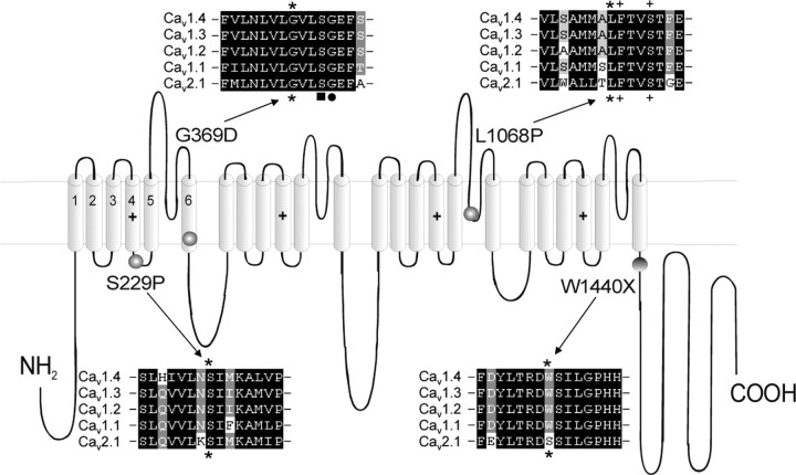
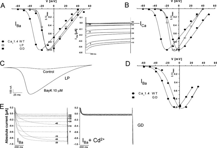
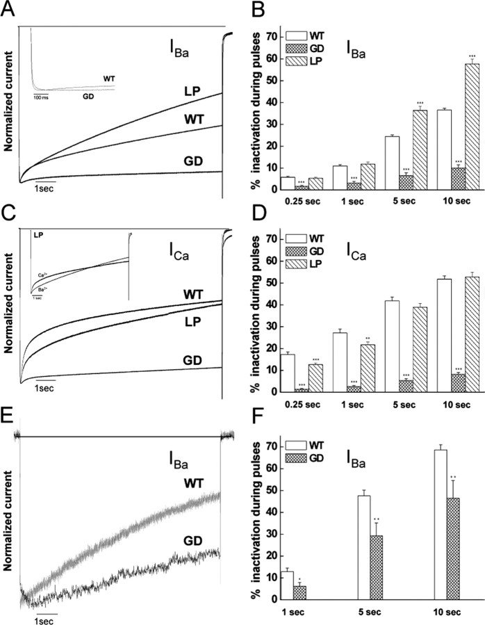
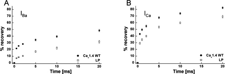
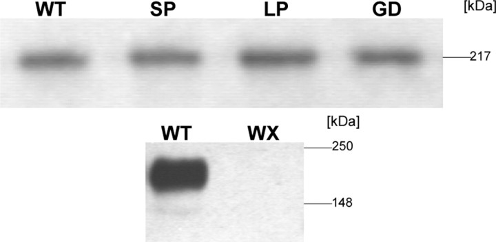
Mutations in the CACNA1F gene (voltage-dependent L-type calcium channel alpha1F subunit) encoding retinal Ca(v)1.4 L-type Ca2+ channels cause X-linked recessive congenital stationary night blindness type 2 (CSNB2). Many of them are predicted to yield nonfunctional channels. Complete loss of Ca(v)1.4 function is therefore regarded as a pathogenetic mechanism for the impaired signaling from photoreceptors to second-order retinal neurons. We investigated the functional consequences of CSNB2 missense mutations S229P, G369D, and L1068P and the C-terminal truncation mutant W1440X. After expression in Xenopus laevis oocytes or human embryonic kidney tsA-201 cells, inward Ca2+ current (I(Ca)) and inward Ba2+ current (I(Ba)) could be recorded from mutations G369D and L1068P. G369D shifted the half-maximal voltage for channel activation (V(0.5,act)) significantly to more negative potentials (>11 mV), slowed inactivation, and removed Ca2+-dependent inactivation. The L1068P mutant yielded currents only in the presence of the channel activator BayK8644. Currents (I(Ba)) inactivated faster than wild type (WT) and recovered more slowly from inactivation (I(Ba) and I(Ca)). No channel activity could be measured for mutants S229P and W1440X after oocyte expression. No W1440X alpha1 protein was detected after expression in tsA-201 cells, whereas S229P (as well as G369D and L1068P) alpha1 subunits were expressed at levels indistinguishable from WT (n = 3). Our data provide unequivocal evidence that CSNB2 missense mutations can induce severe changes in Ca(v)1.4 function, which may decrease (L1068P and S229P) or even increase (G369D) channel activity. The lower activation range of G369D can explain the reduced dynamic range of photoreceptor signaling. Moreover, we demonstrate that loss of channel function of one (L1068P) CSNB2 mutation can be rescued by a Ca2+ channel activator.


Figures

Figure 1.
Figure 1.
Location of missense mutations SP, GD, LP and truncated mutation WX in the Cav1.4α1 subunit. For each mutation, a corresponding ClustalW sequence alignment between human Cav1.4 α1, DHP-sensitive Cav1.3 α1, Cav1.2 α1, Cav1.1 α1, and DHP-insensitive Cav2.1 α1 (NCBI GenBank accession numbers NM_005183, NM_000720, NM_000719, NM_000069, and NM_023035, respectively) was performed in a small region seven amino acids before and after the mutation. All of the amino acids mutated in Cav1.4α1 subunit (indicated by asterisks) were highly conserved among L-type and non-L-type Ca2+ channels (as shown for Cav2.1 α1). In the alignment of mutant LP, two amino acids critical for DHP agonist binding are indicated by + (Yamaguchi et al., 2003). In the alignment of mutant GD, the black square indicates a serine residue important for slow inactivation in IS6 (Shi and Soldatov, 2002), and the black dot indicates the recently found Cav1.2 G406R missense mutation responsible for Timothy syndrome (Splawski et al., 2004).
Figure 2.
Figure 2.
Activation properties of Cav1.4 WT and mutant α1 subunits. A, B, Normalized I-V curves for Cav1.4 WT and mutants GD and LP α1 subunits coexpressed with β3 and α2δ1 subunits in X. laevis oocytes. Ba2+ (A) or Ca2+ (B) were used as charge carriers with 10 μm BayK present in the bath. Representative I-V relationships are shown. For statistics, see Table 1. B, Inset, A representative original trace (n > 3) of Cav1.4 WT Ca2+ currents elicited by depolarizing pulses of 2 sec from -80 to +60 mV. C, Voltage ramps applied from a holding potential of -80 to +70 mV within 360 msec in the absence (control) and presence of 10 μm BayK in the same mutant LP-expressing oocyte. Such a BayK requirement for LP function was observed in all of the experiments (n = 18). A representative experiment is shown. D, Normalized I-V curves for Cav1.4 WT and mutant GD α1 subunits coexpressed with β3 and α2δ1 subunits in tsA-201 cells (as described in Materials and Methods) using 15 mm Ba2+ as charge carrier. Data points are means ± SE (n = 40 for WT and n = 6 for GD). E, Representative family of mutant GD Ba2+ currents elicited by 1 sec depolarizations from -80 to +60 mV before (left) and after (right) the addition of 1 mm Cd2+, which resulted in a total block of the current.
Figure 3.
Figure 3.
Inactivation properties of Cav1.4 WT and mutant α1 subunits. IBa (A, B) and ICa (C, D) through Cav1.4 WT and mutants GD and LP α1 subunits coexpressed with β3 and α2δ1 subunits in X. laevis oocytes were elicited by 10 sec depolarizing pulses from a holding potential of -80 mV to Vmax plus 10 mV (10 μm BayK present). Traces were normalized to the peak current amplitudes. A, C, Representative current traces. B, D, Percentages of current inactivation were calculated after 0.25, 1, 5, and 10 sec depolarizations to the voltage of peak current plus 10 mV. Statistical differences for the percentage of inactivated current were determined by using one-way ANOVA, followed by the Bonferroni multiple-comparison test. Asterisks indicate statistically significant difference to WT (**p < 0.01; ***p < 0.001). GD increased the time-to-peak current (A, inset; WT, 19.5 ± 1.4 msec, n = 23; GD, 68.9 ± 13.3 msec, n = 13; p < 0.001). B, Inset, The complex inactivation time courses of IBa and ICa for mutant LP. E, F, IBa through Cav1.4 WT and mutant GD α1 subunits coexpressed with β3 and α2δ1 subunits in tsA201 cells were elicited by 10 sec depolarizing pulses from a holding potential of -90 mV to Vmax. One representative experiment of six is shown. Currents were normalized to peak IBa. Percentages of current inactivation (F) were measured after 1, 5, and 10 sec depolarizations from -90 mV to the voltage of peak current. Asterisks indicate statistically significant difference (*p < 0.05; **p < 0.01; unpaired Student's t test).
Figure 4.
Figure 4.
Recovery of inactivation of Cav1.4 WT and mutant LP α1 subunits. The percentage of recovery from the inactivated current was determined after 0.5, 1, 2, 5, 10, and 20 sec after a 10 sec depolarization to Vmax plus 10 mV for Cav1.4 WT and mutant LP α1 subunits coexpressed with β3 and α2δ1 subunits in X. laevis oocytes. A concentration of 10 mm Ba2+ (A) or 10 mm Ca2+ (B) was used as charge carriers (10 μm of BayK present). The extent of recovery was statistically significant after all of the time points indicated for IBa and ICa compared with WT (p < 0.001; unpaired Student's t test).
Figure 5.
Figure 5.
Heterologous expression of Cav1.4 WT and different mutant α1 subunits in tsA-201 cells. Human embryonic kidney tsA201 cells were transfected with Cav1.4 WT and mutant GD, LP, SP, and WX α1 subunits together with β3 and α2δ1 subunit cDNAs, as described in Materials and Methods. Cav1.4 α1 subunit proteins were quantified in immunoblots of microsomal membranes prepared from lysed cells after separation on 8% SDS-PAGE gels (10 μg of membrane protein per lane) using a generic anti-α1 sequence-directed antibody (anti-Cav1.1 α1-1382-1400). Cav1.4 WT and mutants migrated with the expected molecular weight of 219 kDa. Staining was absent for the WX mutant (calculated molecular weight of 161 kDa). One representative of three experiments yielding similar results is shown.

References

    1. Ball SL, Powers PA, Shin HS, Morgans CW, Peachey NS, Gregg RG (2002) Role of the β2 subunit of voltage-dependent calcium channels in the retinal outer plexiform layer. Invest Ophthalmol Vis Sci 43: 1595-1603. - PubMed
    1. Barnes S, Kelly ME (2002) Calcium channels at the photoreceptor synapse. Adv Exp Med Biol 514: 465-476. - PubMed
    1. Baumann L, Gerstner A, Zong X, Biel M, Wahl-Schott C (2004) Functional characterization of the L-type Ca2+ channel Cav1.4 α1 from mouse retina. Invest Ophthalmol Vis Sci 45: 708-713. - PubMed
    1. Bech-Hansen NT, Naylor MJ, Maybaum TA, Pearce WG, Koop B, Fishman GA, Mets M, Musarella MA, Boycott KM (1998) Loss-of-function mutations in a calcium-channel α1-subunit gene in Xp11.23 cause incomplete X-linked congenital stationary night blindness. Nat Genet 19: 264-267. - PubMed
    1. Boycott KM, Pearce WG, Bech-Hansen NT (2000) Clinical variability among patients with incomplete X-linked congenital stationary night blindness and a founder mutation in CACNA1F. Can J Ophthalmol 35: 204-213. - PubMed

Publication types

Substances