. 2005 Jan;43(1):199-207.
doi: 10.1128/JCM.43.1.199-207.2005.

Microbial DNA typing by automated repetitive-sequence-based PCR

Affiliations

Microbial DNA typing by automated repetitive-sequence-based PCR

Mimi Healy et al. J Clin Microbiol. 2005 Jan.

Abstract

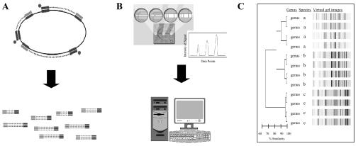
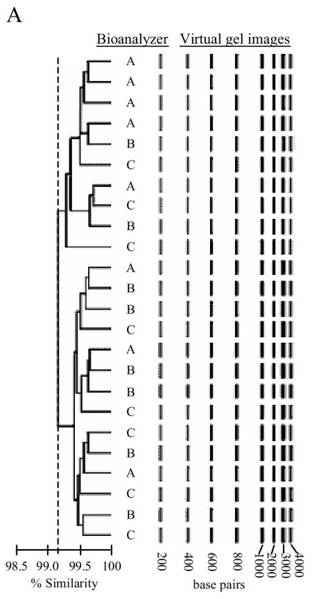
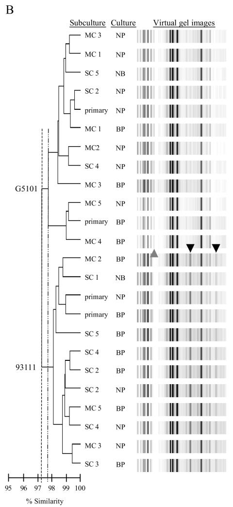
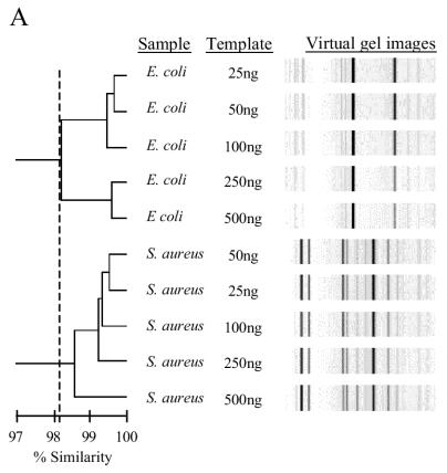
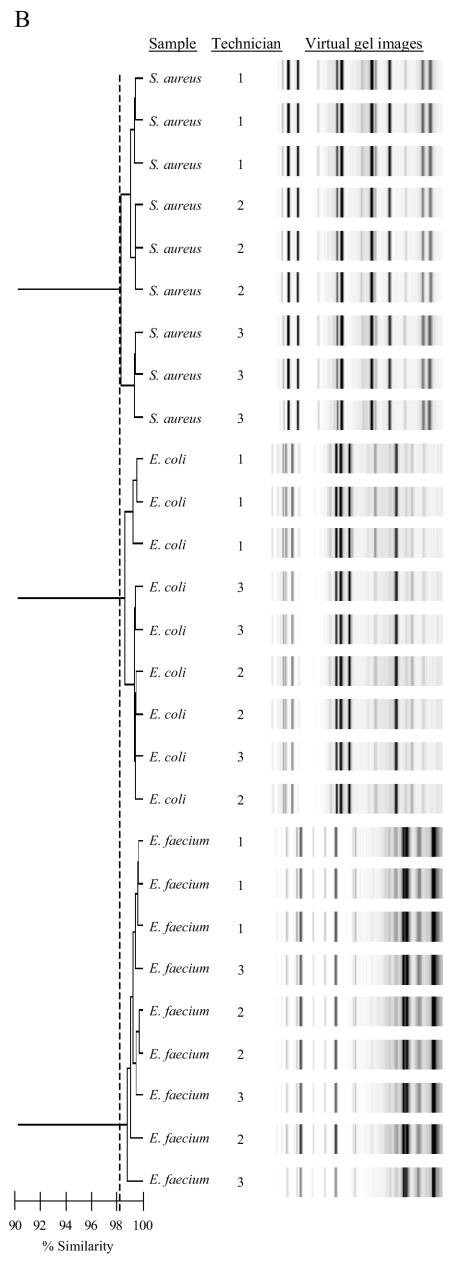
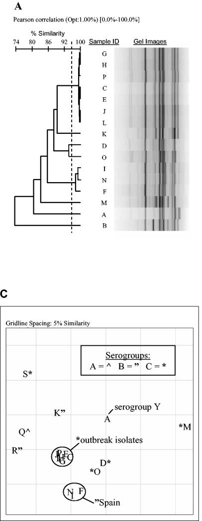
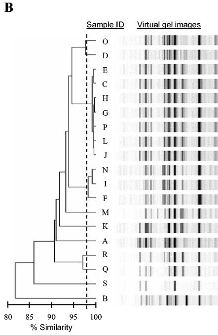
Repetitive sequence-based PCR (rep-PCR) has been recognized as an effective method for bacterial strain typing. Recently, rep-PCR has been commercially adapted to an automated format known as the DiversiLab system to provide a reliable PCR-based typing system for clinical laboratories. We describe the adaptations made to automate rep-PCR and explore the performance and reproducibility of the system as a molecular genotyping tool for bacterial strain typing. The modifications for automation included changes in rep-PCR chemistry and thermal cycling parameters, incorporation of microfluidics-based DNA amplicon fractionation and detection, and Internet-based computer-assisted analysis, reporting, and data storage. The performance and reproducibility of the automated rep-PCR were examined by performing DNA typing and replicate testing with multiple laboratories, personnel, instruments, DNA template concentrations, and culture conditions prior to DNA isolation. Finally, we demonstrated the use of automated rep-PCR for clinical laboratory applications by using isolates from an outbreak of Neisseria meningitidis infections. N. meningitidis outbreak-related strains were distinguished from other isolates. The DiversiLab system is a highly integrated, convenient, and rapid testing platform that may allow clinical laboratories to realize the potential of microbial DNA typing.


Figures

FIG. 1.
FIG. 1.
Schematic diagram of DiversiLab system work flow. (A) rep-PCR primer binds to genomic DNA at multiple sites and allows PCR generation of amplicons of various sizes. (B) The amplicons are separated by microfluidics with the DNA LabChip device, and data are automatically collected and analyzed with the DiversiLab software. (C) A final report is then generated.
FIG. 2.
FIG. 2.
rep-PCR fragment detection reproducibility and pattern stability. (A) Dendrogram and virtual gel images representing a DNA ladder analyzed by three operators using three separate DiversiLab Systems (A to C) during multiple days. (B) Dendrogram and virtual gel images representing rep-PCR fingerprint patterns of E. coli isolates (strains G5101 and 93111) cultured under different growth conditions and typed with the automated rep-PCR system. primary, primary culture from freezer stock; SC, single colony used in subsequent culturing; MC, multiple colony used in subsequent culturing; BP, blood plate; NP, nutrient plate; NB, nutrient broth. The numbers indicate the subculturing step. Arrowheads represent specific PCR amplicons.
FIG. 2.
FIG. 2.
rep-PCR fragment detection reproducibility and pattern stability. (A) Dendrogram and virtual gel images representing a DNA ladder analyzed by three operators using three separate DiversiLab Systems (A to C) during multiple days. (B) Dendrogram and virtual gel images representing rep-PCR fingerprint patterns of E. coli isolates (strains G5101 and 93111) cultured under different growth conditions and typed with the automated rep-PCR system. primary, primary culture from freezer stock; SC, single colony used in subsequent culturing; MC, multiple colony used in subsequent culturing; BP, blood plate; NP, nutrient plate; NB, nutrient broth. The numbers indicate the subculturing step. Arrowheads represent specific PCR amplicons.
FIG. 3.
FIG. 3.
Genomic DNA concentration and technical reproducibility of the patterns generated by automated rep-PCR. (A) Dendrogram and virtual gel images illustrating the effects of genomic DNA concentrations (100, 250, and 500 ng) on rep-PCR fingerprint patterns. (B) Dendrogram and virtual gel images representing rep-PCR fingerprint patterns from E. coli (93111), E. faecium, and S. aureus isolates processed by three laboratory personnel using three DiversiLab systems.
FIG. 3.
FIG. 3.
Genomic DNA concentration and technical reproducibility of the patterns generated by automated rep-PCR. (A) Dendrogram and virtual gel images illustrating the effects of genomic DNA concentrations (100, 250, and 500 ng) on rep-PCR fingerprint patterns. (B) Dendrogram and virtual gel images representing rep-PCR fingerprint patterns from E. coli (93111), E. faecium, and S. aureus isolates processed by three laboratory personnel using three DiversiLab systems.
FIG. 4.
FIG. 4.
DNA fingerprint analysis of rep-PCR with BioNumerics and DiversiLab software of N. meningitidis isolates. (A) Dendrogram analysis and virtual gel image of rep-PCR fingerprint patterns of N. meningitidis isolates (samples A to P) with BioNumerics. (B) Dendrogram analysis and virtual gel images of rep-PCR fingerprint patterns of the N. meningitidis isolates (samples A to S) with DiversiLab software, version 2.1.66. (C) Scatter plot of the N. meningitidis isolates (samples A to S) with the DiversiLab software, version 2.1.66. Isolates C, E, G, H, J, L, and P were obtained from a single outbreak of disease.
FIG. 4.
FIG. 4.
DNA fingerprint analysis of rep-PCR with BioNumerics and DiversiLab software of N. meningitidis isolates. (A) Dendrogram analysis and virtual gel image of rep-PCR fingerprint patterns of N. meningitidis isolates (samples A to P) with BioNumerics. (B) Dendrogram analysis and virtual gel images of rep-PCR fingerprint patterns of the N. meningitidis isolates (samples A to S) with DiversiLab software, version 2.1.66. (C) Scatter plot of the N. meningitidis isolates (samples A to S) with the DiversiLab software, version 2.1.66. Isolates C, E, G, H, J, L, and P were obtained from a single outbreak of disease.

References

    1. Borgdorff, M. W., A. Kolk, D. van Soolingen, J. W. van der Meer, and T. H. Ottenhoff. 2003. Research into new methods for diagnosing, treating and preventing tuberculosis. Ned. Tijdschr. Geneeskd. 147:1838-1841. - PubMed
    1. Cangelosi, G. A., R. J. Freeman, K. N. Lewis, D. Livingston-Rosanoff, K. S. Shah, S. J. Milan, and S. V. Goldberg. 2004. Evaluation of a high-throughput repetitive-sequence-based PCR system for DNA fingerprinting of Mycobacterium tuberculosis and Mycobacterium avium complex strains. J. Clin. Microbiol. 42:2685-2693. - PMC - PubMed
    1. Chau, A. S., C. A. Mendrick, F. J. Sabatelli, D. Loebenberg, and P. M. McNicholas. 2004. Application of real-time quantitative PCR to molecular analysis of Candida albicans strains exhibiting reduced susceptibility to azoles. Antimicrob. Agents Chemother. 48:2124-2131. - PMC - PubMed
    1. Clementino, M. M., I. de Filippis, C. R. Nascimento, R. Branquinho, C. L. Rocha, and O. B. Martins. 2001. PCR analyses of tRNA intergenic spacer, 16S-23S internal transcribed spacer, and randomly amplified polymorphic DNA reveal inter- and intraspecific relationships of Enterobacter cloacae strains. J. Clin. Microbiol. 39:3865-3870. - PMC - PubMed
    1. Cousins, D. V., R. A. Skuce, R. R. Kazwala, J. D. van Embden, et al. 1998. Towards a standardized approach to DNA fingerprinting of Mycobacterium bovis. Int. J. Tuberc. Lung Dis. 2:471-478. - PubMed

Publication types

MeSH terms

LinkOut - more resources