. 2005 Mar;16(3):1282-95.
doi: 10.1091/mbc.e04-07-0578. Epub 2005 Jan 5.

A plasma membrane pool of phosphatidylinositol 4-phosphate is generated by phosphatidylinositol 4-kinase type-III alpha: studies with the PH domains of the oxysterol binding protein and FAPP1

Affiliations

A plasma membrane pool of phosphatidylinositol 4-phosphate is generated by phosphatidylinositol 4-kinase type-III alpha: studies with the PH domains of the oxysterol binding protein and FAPP1

Andras Balla et al. Mol Biol Cell. 2005 Mar.

Abstract

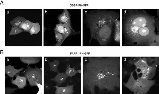
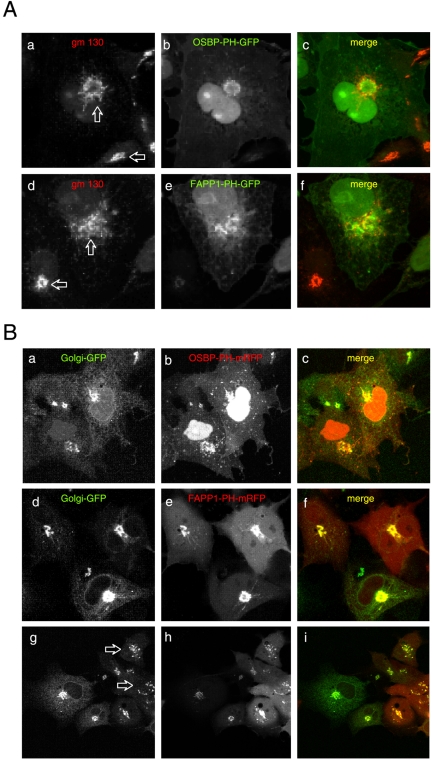
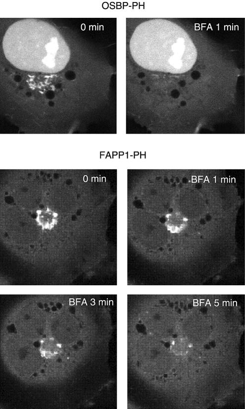
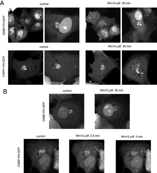
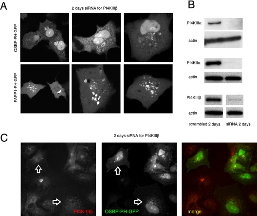
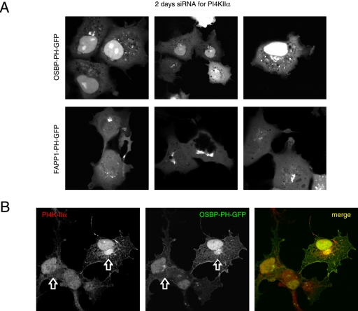
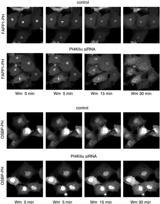
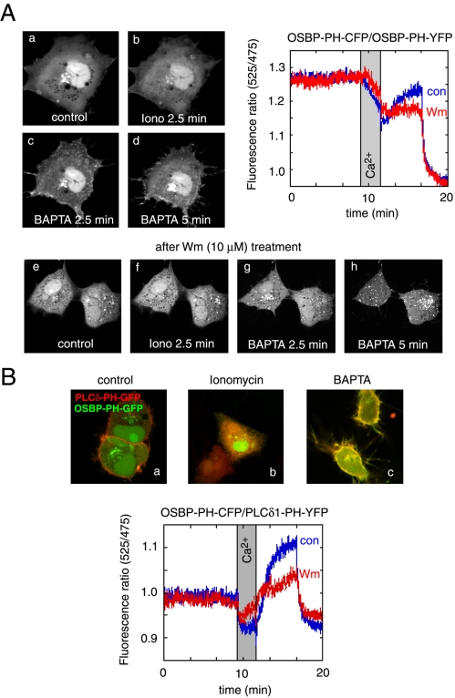
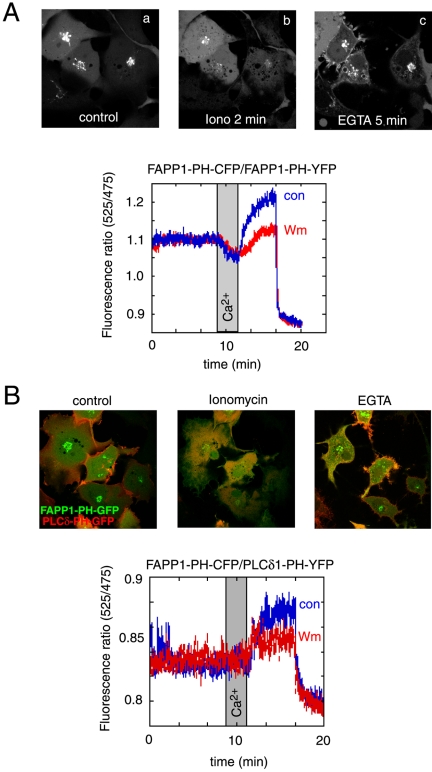
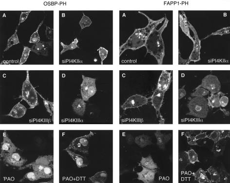
The PH domains of OSBP and FAPP1 fused to GFP were used to monitor PI(4)P distribution in COS-7 cells during manipulations of PI 4-kinase (PI4K) activities. Both domains were associated with the Golgi and small cytoplasmic vesicles, and a small fraction of OSBP-PH was found at the plasma membrane (PM). Inhibition of type-III PI4Ks with 10 microM wortmannin (Wm) significantly reduced but did not abolish Golgi localization of either PH domains. Downregulation of PI4KIIalpha or PI4KIIIbeta by siRNA reduced the localization of the PH domains to the Golgi and in the former case any remaining Golgi localization was eliminated by Wm treatment. PLC activation by Ca2+ ionophores dissociated the domains from all membranes, but after Ca2+ chelation, they rapidly reassociated with the Golgi, the intracellular vesicles and with the PM. PM association of the domains was significantly higher after the Ca2+ transient and was abolished by Wm pretreatment. PM relocalization was not affected by down-regulation of PI4KIIIbeta or -IIalpha, but was inhibited by down-regulation of PI4KIIIalpha, or by 10 microM PAO, which also inhibits PI4KIIIalpha. Our data suggest that these PH domains detect PI(4)P formation in extra-Golgi compartments under dynamic conditions and that various PI4Ks regulate PI(4)P synthesis in distinct cellular compartments.


Figures

Figure 1.
Figure 1.
Cellular distribution of OSBP-PH-GFP (A) and FAPP1-PH-GFP (B) expressed in COS-7 cells. Cells were transiently transfected with the indicated PH domain-GFP chimera for 24 h, and live cells were studied by confocal microscopy at 35°C. In most cells showing low-to-moderate expression levels both constructs show localization to the Golgi compartment (a and b panels in both A and B). In many cells expressing low levels of either construct dynamic tubular structures emanate from the Golgi, reminiscent of the effects of early BFA treatment (panels c). At higher expression levels, OSBP-PH showed several small vesicles around the Golgi area and also in more peripheral locations, whereas the FAPP1-PH domain generated multiple larger vesicles strongly positive for the domain in their limiting membranes (panels d on A and B, respectively). Notable difference is the nuclear accumulation of OSBP-PH but not FAPP1-PH, especially in cells expressing the protein at higher levels.
Figure 2.
Figure 2.
Effects of overexpressed OSBP- and FAPP1-PH-GFP on the distribution of Golgi markers. COS-7 cells were transfected for 24 h with either the GFP-fused (A) or the mRFP-fused (B) respective PH domains and studied either as fixed cells immunostained for the cis-Golgi marker, gm130 (A) or cotransfected with a GFP construct containing the N-terminal transmembrane domain of the β1,4-galactosyl transferase enzyme (Golgi-GFP) and observed live (B). Note the fragmentation and tubulation of the cis-Golgi marker in the cells expressing higher levels of either PH domains (compare the structures indicated by the two arrows on A) and the loss of tight colocalization observed in cells expressing the constructs at low level. (B) Similar fragmentation of the Golgi was observed in live cells expressing high levels of the FAPP1-PH-mRFP (arrows in bottom row), but this effect was not so prominent with the OSBP-PH-mRFP (top row).
Figure 3.
Figure 3.
Effects of brefeldin A (BFA) treatment on the localization of OSBP- and FAPP1-PH-GFP in COS-7 cells. Cells were transfected with the indicated constructs and live cells were examined by laser confocal microscopy 24 h later at 35°C. (A) After addition of BFA (5 μg/ml) OSBP-PH-GFP dissociated from the Golgi very rapidly (within 1 min), whereas the FAPP1-PH domain required a longer period (3–5 min).
Figure 4.
Figure 4.
Effects of wortmannin (Wm) treatment on the localization of the OSBP or FAPP1-PH-GFP fusion proteins in COS-7 cells. COS-7 cells were transfected with the indicated PH-GFP construct for 24 h and studied as live cells by confocal microscopy. (A) Cells were treated with 10 μM Wm for 30 min (at 37°C) to fully inhibit the type-III PI4Ks. There was a decrease in the Golgi localization of both PH domains after Wm treatment, but there was still a significant localization in vesicular compartments, often associated with the Golgi area. In most cases these vesicles were more dispersed than those in untreated cells. (B) The difference between pre- and post-Wm treatment is illustrated in a cell that was recorded both before and after the Wm treatment. The bottom panel shows a cell that rapidly loses some of its OSBP-PH domain localization after Wm treatment. Figure 7 shows additional examples of the effects of Wm treatment on the Golgi localization.
Figure 5.
Figure 5.
Effects of down-regulation of the type-IIIβ PI4K by siRNA treatment on the localization of OSBP- and FAPP1-PH-GFP in live (A) and OSBP-PH-GFP in fixed (C) COS-7 cells. COS-7 cells were treated with double-stranded RNA designed to interfere with expression of PI4KIIIβ for 2 consecutive days and transfected with the PH-GFP constructs on the third day for an additional day as described in Materials and Methods. Cells were either studied live at 35 C (A) or analyzed after fixation and immunostaining with a polyclonal anti-PI4KIIIβ antibody (C). The arrows point to two cells in which PI4KIIIβ is largely eliminated. To determine the effect of siRNA treatment, cells treated identically with the various siRNAs directed against the three PI4K enzymes were subjected to Western blot analysis using antibodies against the respective kinases and the blots were also analyzed for actin (B). In the picture showing the effect of PI4KIIIβ knockdown the control and siRNA-treated samples were not loaded on adjacent lanes, indicated by the gap between the images. Nevertheless the images are derived from the same gel scanned and processed with identical settings.
Figure 6.
Figure 6.
Effects of down-regulation of the type-IIα PI4K by siRNA treatment on the localization of OSBP- and FAPP1-PH-GFP in live (A) and OSBP-PH-GFP in fixed (B) COS-7 cells. COS-7 cells were treated with double-stranded RNA designed to interfere with expression of PI4KIIα for 2 consecutive days and transfected with the PH-GFP constructs for an additional day as described in Materials and Methods. Cells were either studied live at 35°C (A) or analyzed after fixation and immunostaining with a polyclonal anti-PI4KIIα antibody (B; Guo et al., 2003). The arrows point to the Golgi area of two cells, one in which the PI4KIIα is largely eliminated and another in which it is still readily detectable.
Figure 7.
Figure 7.
Effect of wortmannin treatment on the localization of the OSBP- and FAPP1-PH-GFP after 2-d treatment with siRNA against PI4KIIα. COS-7 cells were treated with siRNA and transfected with the PH domain constructs as described in the legend to Figure 5. Live cells were studied at 35°C and treated with 10 μM Wm for the indicated times. Note the complete loss of localization of the PH domains in the siRNA-treated cells but not in the controls.
Figure 8.
Figure 8.
Effects of cytoplasmic [Ca2+] increases on the distribution of the OSBP-PH-GFP fusion protein. Cells were transfected with the indicated constructs and live cells were examined by confocal microscopy (Zeiss LSM410) at 35°C. (A) Addition of ionomycin (10 μM) in the presence of 2 mM external Ca2+ caused the translocation of the PH domain from the Golgi to the cytoplasm. Chelation of Ca2+ by BAPTA restores the localization in the Golgi and induces a prominent plasma membrane localization of the PH domain. Wortmannin (Wm) pretreatment prevents the plasma membrane localization but not the vesicular localization of the PH domain after ionomycin/BAPTA treatment (A, lower series). FRET measurements between CFP- and YFP-fused OSBP-PH domains coexpressed in COS-7 cells show a high initial FRET value (probably because of the high nuclear accumulation of the constructs), which decreases after ionomycin addition, but shows a rapid increase after Ca2+ chelation, which is mostly sensitive to Wm (10 μM) treatment (A, graph). (B) Simultaneous monitoring of the OSBP-PH and PLCδ1-PH domain translocations by coexpression of GFP and mRFP fused PH domains, respectively. Note the prominent colocalization of the red and green signals after (but not before) the iono/BAPTA treatment. FRET measurements between the same domains fused to CFP and YFP (OSBP-PH-CFP and PLCδ1-PH-YFP) shows a small initial FRET that is abolished after ionomycin treatment and returns well above baseline after Ca2+ chelation. This FRET analysis shows the plasma membrane component of the OSBP-PH movement that is significantly reduced after Wm treatment.
Figure 9.
Figure 9.
Effects of cytoplasmic [Ca2+] increases on the distribution of the FAPP1-PH-GFP fusion protein. See legend to Figure 8 for experimental details. (A) Addition of ionomycin (10 μM) in the presence of 2 mM external Ca2+ caused the translocation of the PH domain from the Golgi to the cytoplasm. Chelation of Ca2+ by EGTA restores the localization in the Golgi but the plasma membrane localization of the FAPP1-PH domain is less prominent that that of the OSBP-PH. FRET measurements between CFP- and YFP-fused FAPP1-PH domains coexpressed in COS-7 cells show a rapid decrease after ionomycin addition and show a rapid increase above basal after Ca2+ chelation. About 50% of this increase is sensitive to Wm (10 μM) treatment (A, graph). (B) Simultaneous monitoring of the FAPP1-PH and PLCδ1-PH domain translocations by coexpression of GFP and mRFP fused PH domains, respectively. The colocalization of the red and green signals after the iono/EGTA treatment is not as prominent as with the OSBP-PH domain. FRET measurements between the same domains fused to CFP and YFP (FAPP1-PH-CFP and PLCδ1-PH-YFP) shows no initial FRET and no change after ionomycin treatment. FRET values; however, increase above baseline after Ca2+ chelation. This FRET analysis shows the plasma membrane component of the FAPP1-PH movement that is smaller than with the OSBP-PH and is also significantly reduced after Wm treatment.
Figure 10.
Figure 10.
Relocalization of OSBP- and FAPP1PH-GFP in COS-7 cells in which PI4K isoforms were down-regulated or inhibited. Cells were treated with siRNA and transfected with the plasmid DNA as described in the legend to Figure 5. Cells were studied live on the temperature-controlled stage of a Zeiss 410 laser confocal microscope at 35°C. Cells were stimulated with ionomycin (10 μM) until most of their localized PH domains were released from the Golgi (2–3 min), at which point Ca2+ was chelated by the addition of EGTA (7 mM) and images were recorded after 3–7 min. When indicated, PAO (10 μM) was added 10 min before ionomycin either in the presence or absence of 1 mM DTT. Note that the prominent plasma membrane localization of both PH domains was abolished in cells treated with PAO or in which the PI4KIIIα was down-regulated.

References

    1. Audhya, A., Foti, M., and Emr, S. D. (2000). Distinct roles for the yeast phosphatidylinositol 4-kinases, stt4p and pik1p, in secretion, cell growth, and organelle membrane dynamics. Mol. Biol. Cell 11, 2673-2689. - PMC - PubMed
    1. Audhya, A., and Emr, S. D. (2002). Stt4 PI 4-kinase localizes to the plasma membrane and functions in the Pkc1-mediated MAP kinase cascade. Dev. Cell 2, 593-605. - PubMed
    1. Balla, A., Tuymetova, G., Barshishat, M., Geiszt, M., and Balla, T. (2002). Characterization of type II phosphatidylinositol 4-kinase isoforms reveals association of the enzymes with endosomal vesicular compartments. J. Biol. Chem. 277, 20041-20050. - PubMed
    1. Balla, T., Bondeva, T., and Varnai, P. (2000). How accurately can we image inositol lipids in live cells? Trends Pharmacol. Sci. 21, 238-241. - PubMed
    1. Balla, T. and Varnai, P. (2002). Visualizing cellular phosphoinositide pools with GFP-fused protein-modules. Sci. STKE 125, PL3, 1-16. - PubMed

Publication types

MeSH terms

Substances