. 2005 Jan 26;24(2):270-82.
doi: 10.1038/sj.emboj.7600530. Epub 2005 Jan 6.

Bacterial chromosome segregation: structure and DNA binding of the Soj dimer--a conserved biological switch

Affiliations

Bacterial chromosome segregation: structure and DNA binding of the Soj dimer--a conserved biological switch

Thomas A Leonard et al. EMBO J. .

Abstract

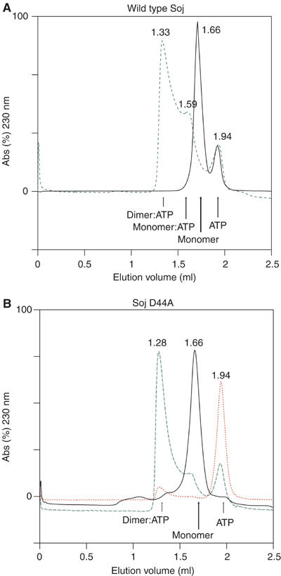
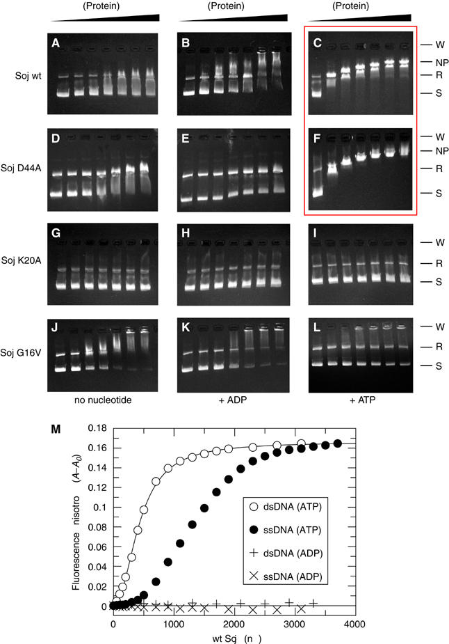
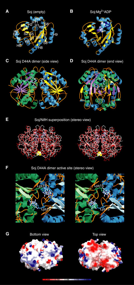
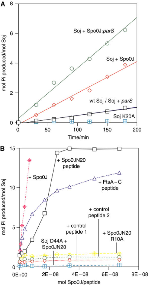
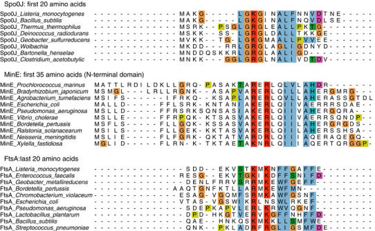
Soj and Spo0J of the Gram-negative hyperthermophile Thermus thermophilus belong to the conserved ParAB family of bacterial proteins implicated in plasmid and chromosome partitioning. Spo0J binds to DNA near the replication origin and localises at the poles following initiation of replication. Soj oscillates in the nucleoid region in an ATP- and Spo0J-dependent fashion. Here, we show that Soj undergoes ATP-dependent dimerisation in solution and forms nucleoprotein filaments with DNA. Crystal structures of Soj in three nucleotide states demonstrate that the empty and ADP-bound states are monomeric, while a hydrolysis-deficient mutant, D44A, is capable of forming a nucleotide 'sandwich' dimer. Soj ATPase activity is stimulated by Spo0J or the N-terminal 20 amino-acid peptide of Spo0J. Our analysis shows that dimerisation and activation involving a peptide containing a Lys/Arg is conserved for Soj, ParA and MinD and their modulators Spo0J, ParB and MinE, respectively. By homology to the nitrogenase iron protein and the GTPases Ffh/FtsY, we suggest that Soj dimerisation and regulation represent a conserved biological switch.


Figures

Figure 1
Figure 1
Size exclusion chromatography of T. thermophilus wild-type Soj and Soj D44A in the presence and absence of ATP. The elution volume of each species is indicated in millilitres above the respective trace. (A) In the absence of ATP, wild-type Soj elutes as a monomeric species (black). In the presence of ATP, Soj elutes as a dimer–monomer equilibrium (green). (B) Soj D44A, deficient in nucleotide hydrolysis, also elutes as a monomer in the absence of ATP (black), but elutes almost solely as a dimer in the presence of ATP (green). Red: absorbance at 260 nm indicating the elution volume of ATP and the presence of ATP in the dimeric protein.
Figure 2
Figure 2
Sedimentation velocity analysis of wild-type Soj (A, E), Soj K20A (B, F), Soj D44A (C, G) and Soj G16V (D, H), without and with ATP, respectively. For each sample, a series of scans (at intervals of ∼15 min) are shown. Also shown are the residuals from fitting a series of 12 scans (at intervals of ∼1.5 min), near the middle of the sedimentation run, to a model for either one component (most runs) or two components specifically for mutant D44A+ATP. This fitting was used to estimate the sedimentation and diffusion coefficients, and hence calculate the molecular mass, for each component and the random distributions of the residuals suggest that the fits are valid.
Figure 3
Figure 3
Electrophoretic mobility shift assays of Soj DNA binding. Wild-type Soj, hydrolysis-deficient Soj D44A, nucleotide-binding-deficient Soj K20A and dimerisation-deficient Soj G16V bind to DNA only weakly at saturating protein concentrations in the absence of nucleotide (A, D, G, J, left) and in the presence of ADP (B, E, H, K, middle). Soj K20A and Soj G16V fail to bind DNA in the presence of ATP (I, L), indicating that ATP-dependent dimerisation is necessary for DNA binding. Wild-type Soj and Soj D44A shift DNA in a concentration-dependent fashion until saturation in the presence of ATP (C, F). Hydrolysis of ATP by wild-type Soj results in the presence of multiple shifted bands of various molecular weights (C), while hydrolysis-deficient Soj D44A produces a single shifted band at all protein concentrations (F). Key: W=wells, NP=nucleoprotein filament, R=relaxed, S=supercoiled. (M) Fluorescence anisotropy analysis of ATP-dependent Soj binding to DNA.
Figure 4
Figure 4
DNA-dependent pelleting of Soj. Samples were performed in duplicates for reliability. Wild-type Soj is found exclusively in the supernatant when centrifuged in the presence of DNA but without ATP. In the presence of ATP, a substantial fraction (one-third) of wild-type Soj is found in the pellet. Hydrolysis-deficient Soj D44A is also found in the pellet in the presence of ATP and DNA, but to a greater extent than wild-type Soj (experiments performed at above saturating protein concentrations: two Soj dimers: one 24 bp binding site), indicating that ATP hydrolysis results in dissociation from the DNA. These results indicate that ATP-dependent dimerisation is a prerequisite for DNA binding.
Figure 5
Figure 5
Electron micrographs of Soj nucleoprotein filaments. (A) Wild-type Soj+plasmid DNA (pU0J, a 2938 bp pUC19 derivative) in the absence of nucleotide. (B) Wild-type Soj+pU0J+ADP. (C) Wild-type Soj+linearised pU0J+ATP produces nucleoprotein filaments which are indistinguishable from those produced by (D) Soj D44A under the same conditions. (E) Nucleoprotein filaments formed by Soj D44A in the presence of relaxed, open circle pU0J+ATP. (FH) High-magnification images of single nucleoprotein filaments, indicating a regular, perhaps helical arrangement.
Figure 6
Figure 6
Crystal structures of Soj. (A) Crystal structure of Soj in the empty state at 1.6 Å. The arrangement of the sheet and helices follows that of the MinD family of ATPases (Cordell and Löwe, 2001; Hayashi et al, 2001; Sakai et al, 2001). (B) Structure of Soj:Mg2+ADP at 2.1 Å. Nucleotide binding is coupled to rearrangement of the P-loop. (C) Structure of hydrolysis-deficient Soj D44A in the dimeric state (side view), indicating the symmetrical assembly of the two monomers and the close proximity of the two nucleotides. (D) End view of Soj D44A dimer. (E) Structure of Soj D44A superimposed on the structure of nitrogenase iron protein from A. vinelandii (PDB ID: 1n2c) (Schindelin et al, 1997). The high structural homology between the two proteins indicates that the dimeric structure of Soj is correct. (F) Stereo view of the Soj D44A dimer active site. The nucleotide-binding surface of each monomer contributes to the formation of the active site chamber, which accommodates two molecules of ATP. Each monomer also contributes a universally conserved lysine (Lys15), which stabilises the negative charges on the opposing ATP. (G) Bottom and top views of the electrostatic surface potential maps of the Soj D44A dimer. The top view clearly indicates two patches of negative charge (one on each monomer) and, importantly, the existence of a cleft between the two monomers in which the surfaces are entirely complementary.
Figure 7
Figure 7
(A) Time course activation of Soj ATPase activity. Wild-type Soj displays low basal ATPase activity (black squares). Soj is stimulated approximately three-fold by Spo0J (red diamonds) and by almost an order of magnitude by Spo0J in the presence of parS DNA (green circles). Soj K20A is devoid of ATPase activity. (B) Spo0J activation of Soj ATPase activity. Spo0J strongly stimulates Soj ATPase activity at nanomolar concentrations (red hatched diamonds). Soj can also be stimulated by Spo0JN20, a 20 amino-acid peptide from the extreme N-terminus of Spo0J (black squares), although the peptide exhibits only a modest 8% of the activation stimulated by an equimolar amount of full-length Spo0J. Soj is not stimulated by either of the control peptides (red diamonds and green circles) and hydrolysis-deficient Soj D44A is not stimulated by Spo0JN20 (blue hatched squares). The Spo0JN20 R10A peptide, which has the putative catalytic arginine mutated to alanine, fails to stimulate Soj (yellow hatched diamonds), indicating that this residue is critical for activation of ATP hydrolysis. Interestingly, a 19 amino-acid peptide representing the conserved extreme C-terminus of FtsA also strongly stimulates Soj, but to a lesser extent than Spo0JN20. The kinetics of activation by Spo0JN20 and full-length Spo0J indicate possible cooperativity of binding and activation, consistent with dimeric Spo0J binding dimeric Soj.
Figure 8
Figure 8
Sequence alignment of putative activating peptides of Spo0J, MinE and FtsA. Alignment of the N-terminal regions of Spo0J and MinE proteins with the C-terminus of FtsA proteins reveals a remarkable sequence homology, which includes a universally conserved basic (lys/arg) residue (arginine 10 in T. thermophilus Spo0J). The functional homology of these putative activating peptides is further strengthened by the observations that E. coli FtsA C-terminus can stimulate Soj and mutation of the arginine 10 to alanine in the Spo0JN20 peptide abrogates stimulation of ATP hydrolysis (Figure 7).

References

    1. Cervin MA, Spiegelman GB, Raether B, Ohlsen K, Perego M, Hoch JA (1998) A negative regulator linking chromosome segregation to developmental transcription in Bacillus subtilis. Mol Microbiol 29: 85–95 - PubMed
    1. Chan KM, Delfert D, Junger KD (1986) A direct colorimetric assay for Ca2+-stimulated ATPase activity. Anal Biochem 157: 375–380 - PubMed
    1. Cordell SC, Löwe J (2001) Crystal structure of the bacterial cell division regulator MinD. FEBS Lett 492: 160–165 - PubMed
    1. Draper GC, Gober JW (2002) Bacterial chromosome segregation. Annu Rev Microbiol 56: 567–597 - PubMed
    1. Easter J Jr, Gober JW (2002) ParB-stimulated nucleotide exchange regulates a switch in functionally distinct ParA activities. Mol Cell 10: 427–434 - PubMed