. 2005 Jan 26;24(2):325-35.
doi: 10.1038/sj.emboj.7600526. Epub 2005 Jan 6.

Identification of neuromedin S and its possible role in the mammalian circadian oscillator system

Affiliations

Identification of neuromedin S and its possible role in the mammalian circadian oscillator system

Kenji Mori et al. EMBO J. .

Abstract

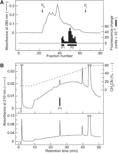
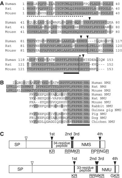
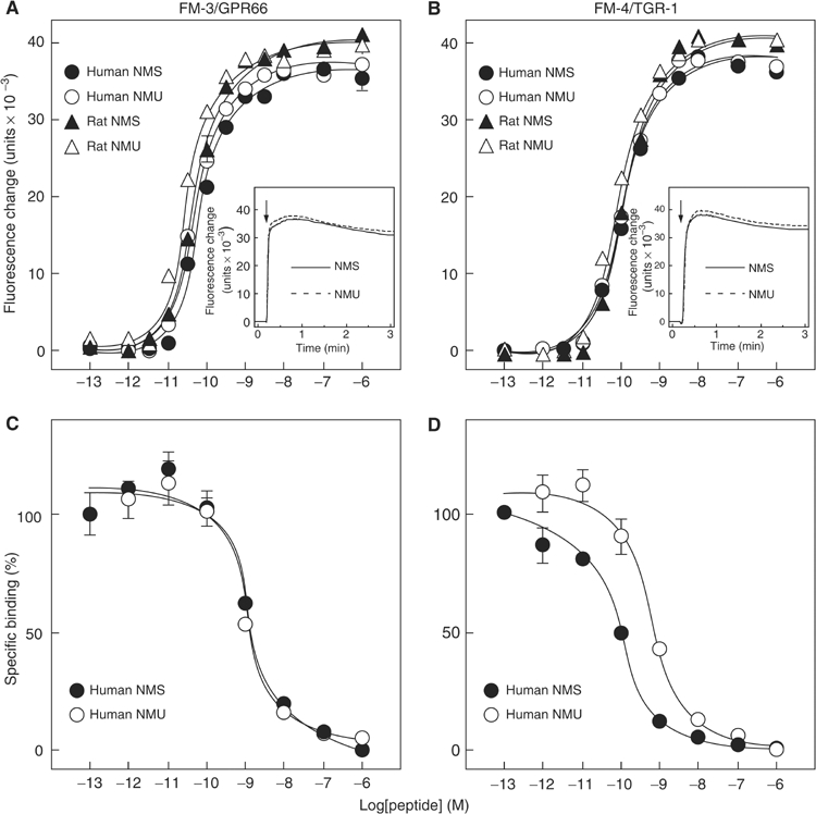
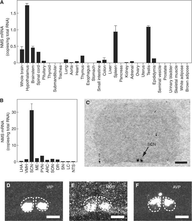
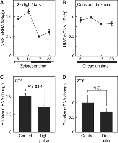
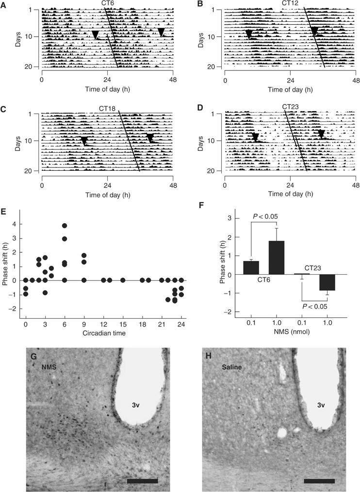
The discovery of neuropeptides has resulted in an increased understanding of novel regulatory mechanisms of certain physiological phenomena. Here we identify a novel neuropeptide of 36 amino-acid residues in rat brain as an endogenous ligand for the orphan G protein-coupled receptor FM-4/TGR-1, which was identified to date as the neuromedin U (NMU) receptor, and designate this peptide 'neuromedin S (NMS)' because it is specifically expressed in the suprachiasmatic nuclei (SCN) of the hypothalamus. NMS shares a C-terminal core structure with NMU. The NMS precursor contains another novel peptide. NMS mRNA is highly expressed in the central nervous system, spleen and testis. In rat brain, NMS expression is restricted to the core of the SCN and has a diurnal peak under light/dark cycling, but remains stable under constant darkness. Intracerebroventricular administration of NMS in rats activates SCN neurons and induces nonphotic type phase shifts in the circadian rhythm of locomotor activity. These findings suggest that NMS in the SCN is implicated in the regulation of circadian rhythms through autocrine and/or paracrine actions.


Figures

Figure 1
Figure 1
Purification of NMS. (A) Gel filtration on a Sephadex G-50 (fine) column of the SP-III fraction from 510 g of rat brain. Black bars indicate fluorescence change due to [Ca2+]i increase in CHO/FM-4. Active fractions containing NMS (P1) and NMU (P2) are indicated. V0, void volume; Vt, total volume. (B) Chromatographic comparison using RP-HPLC of natural NMS (upper panel) and synthetic NMS (lower panel). The black bar indicates [Ca2+]i -increasing activity (upper panel). Each peptide was applied to a Chemcosorb 3ODS-H column. The flow rate was 0.2 ml/min. Solvent system, a linear gradient elution from (A) to (B) for 40 min. H2O:CH3CN:10% TFA for (A) was 90:10:1, for (B) 40:60:1 (v/v).
Figure 2
Figure 2
Structure of NMS. (A) Amino-acid sequences of human, rat and mouse prepro-NMS. Identical residues are shaded. The dotted line denotes the predicted signal peptide. The arrowheads indicate proteolytic processing sites. The asterisk shows a glycine residue, which serves as an amide donor for C-terminal amidation. NMS sequences are boxed. Sequences conserved between NMS and NMU are indicated by a solid underline. The sequence data for the human, rat and mouse NMS cDNAs have been submitted to the DDBJ/EMBL/GenBank databases under accession nos. AB164464, AB164465 and AB164466, respectively. (B) Sequence comparison of NMS and NMU. Human, rat and mouse NMS and NMU sequences are aligned. Residues identical between peptides are shaded. Conserved core sequences are indicated by a solid underline. (C) Schematic representation of the preproproteins of rat NMS and NMU. The preproproteins of NMS and NMU are represented by boxes divided into protein domain, proportional to their length. The open and filled arrowheads indicate cleavage sites by signal peptidase and proprotein convertase, respectively. The sequences of the proteolytic processing sites are shown. The basic amino-acid residues recognized by proprotein convertase are underlined. SP, signal peptide.
Figure 3
Figure 3
Pharmacological characterization of synthetic NMS using human FM-3/GPR66 and FM-4/TGR-1 stably expressed in CHO cells. (A, B) Dose-response relationships of [Ca2+]i change for human NMS (filled circle), human NMU (open circle), rat NMS (filled triangle) and rat NMU (open triangle) in CHO/FM-3 (A) and CHO/FM-4 (B) cells. Data points are means±s.e.m. of triplicates for each experiment. Insets show the time course of [Ca2+]i changes induced by human NMS (solid line) and human NMU (dotted line). Each peptide (10−8 M) was added at the time indicated by the arrow. (C, D) Competitive radioligand binding analysis. [125I-Tyr0]-human NMS binding to FM-3/GPR66 (C) and FM-4/TGR-1 (D) was displaced by increasing concentrations of human NMS (filled circle) and human NMU (open circle). Data were determined in triplicate.
Figure 4
Figure 4
Expression studies of rat NMS. (A, B) Quantitative RT–PCR analysis of the NMS mRNA in a rat multiple-tissue cDNA panel. Each column represents the mean±s.e.m. of triplicate experiments. LHA, lateral hypothalamic area; VMH, ventromedial hypothalamus; SCN, suprachiasmatic nucleus; ME, median eminence; PVN, paraventricular nucleus; ARC, arcuate nucleus; SON, supraoptic nucleus; DR, dorsal raphe; SN, substantia nigra; LC, locus coeruleus; NTS, nucleus of the solitary tract. (C) Autoradiogram of the NMS mRNA expression in a coronal section of the rat brain. Scale bar, 2 mm. (D–F) Distribution of VIP (D), NMS (E) and AVP (F) mRNA in the rat SCN. Serial sections were used. The SCN is indicated with a dotted line. Scale bar, 500 μm.
Figure 5
Figure 5
Expression pattern of rat NMS within the SCN. (A, B) Temporal expression profiles of the NMS mRNA. Animals were maintained under 12-h light/dark cycles (A) or constant darkness for 2 days (B). The amount of NMS mRNA was quantified by in situ hybridization analysis. The experiments were performed side by side. Data represent the means±s.e.m. of 3–4 animals. Open and filled horizontal bars indicate light and dark periods, respectively. (C, D) Responses of NMS expression to light exposure under conditions of constant darkness and to a dark pulse during the light period of a 12-h light/dark cycle. Animals maintained in constant darkness for 2 days were exposed to a light pulse for 30 min at CT6 (C), or animals maintained under 12-h light/dark cycles were exposed to a dark pulse for 30 min at ZT6 (D). Brain samples were collected 1 h after exposure to the light or dark pulse. Data are presented as the means±s.e.m. of 3–4 animals, and are shown as relative changes in NMS mRNA levels.
Figure 6
Figure 6
In vivo experiments with rat NMS. (A–D) Phase shifts of the circadian rhythm of locomotor activity induced by ICV administration of NMS. Representative doubled-plotted actograms of locomotor activity are shown. Rats were ICV administered with rat NMS (1 nmol) at CT6 (A), CT12 (B), CT18 (C) and CT23 (D). Each arrowhead indicates the time of NMS administration. Each line shows the regression lines drawn based on the daily onset of locomotor activity before and after administration. (E) Phase-response plot for ICV administration of 1 nmol NMS. The plus and minus values indicate phase advance and delay, respectively. n=3–5. The data points at CT12, 13, 15, 18 and 19 overlap. (F) Dependence of circadian rhythm phase shift on NMS administration. ICV administration of NMS at CT6 and CT23 induced significant phase advance and delay, respectively, in a dose-dependent manner. n=4–5 per group. (G, H) Induction of c-Fos protein expression within the SCN following ICV administration of NMS. Rats were administered at CT6 with 1 nmol NMS (G) or saline (H). 3v, third ventricle. Scale bar, 150 μm.

References

    1. Barsh GS, Farooqi IS, O'Rahilly S (2000) Genetics of body-weight regulation. Nature 404: 644–651 - PubMed
    1. Brown DR, Quito FL (1988) Neuromedin U octapeptide alters ion transport in porcine jejunum. Eur J Pharmacol 155: 159–162 - PubMed
    1. Castel M, Belenky M, Cohen S, Wagner S, Schwartz WJ (1997) Light-induced c-Fos expression in the mouse suprachiasmatic nucleus: immunoelectron microscopy reveals co-localization in multiple cell types. Eur J Neurosci 9: 1950–1960 - PubMed
    1. Cheng MY, Bullock CM, Li C, Lee AG, Bermak JC, Belluzzi J, Weaver DR, Leslie FM, Zhou QY (2002) Prokineticin 2 transmits the behavioural circadian rhythm of the suprachiasmatic nucleus. Nature 417: 405–410 - PubMed
    1. Chu C, Jin Q, Kunitake T, Kato K, Nabekura T, Nakazato M, Kangawa K, Kannan H (2002) Cardiovascular actions of central neuromedin U in conscious rats. Regul Peptides 105: 29–34 - PubMed

Publication types