. 2005 May 15;388(Pt 1):143-50.
doi: 10.1042/BJ20041623.

IRP1 Ser-711 is a phosphorylation site, critical for regulation of RNA-binding and aconitase activities

Affiliations

IRP1 Ser-711 is a phosphorylation site, critical for regulation of RNA-binding and aconitase activities

Carine Fillebeen et al. Biochem J. .

Abstract

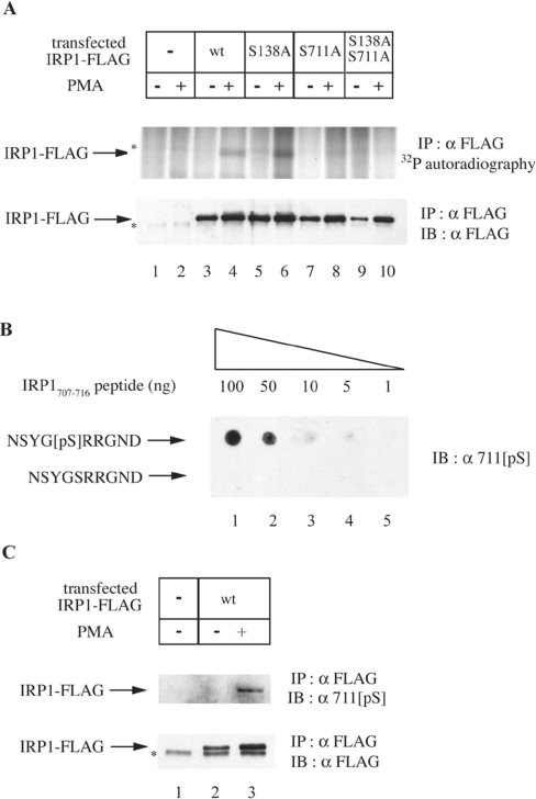
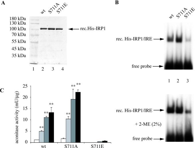
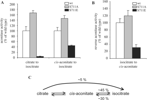
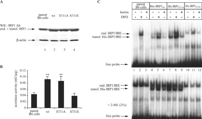
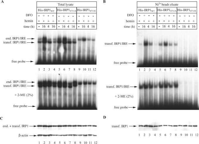
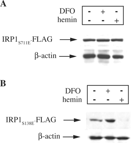
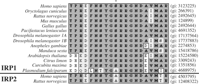
In iron-starved cells, IRP1 (iron regulatory protein 1) binds to mRNA iron-responsive elements and controls their translation or stability. In response to increased iron levels, RNA-binding is inhibited on assembly of a cubane [4Fe-4S] cluster, which renders IRP1 to a cytosolic aconitase. Phosphorylation at conserved serine residues may also regulate the activities of IRP1. We demonstrate that Ser-711 is a phosphorylation site in HEK-293 cells (human embryonic kidney 293 cells) treated with PMA, and we study the effects of the S711E (Ser-711-->Glu) mutation on IRP1 functions. A highly purified preparation of recombinant IRP1(S711E) displays negligible IRE-binding and aconitase activities. It appears that the first step in the aconitase reaction (conversion of citrate into the intermediate cis-aconitate) is more severely affected, as recombinant IRP1(S711E) retains approx. 45% of its capacity to catalyse the conversion of cis-aconitate into the end-product isocitrate. When expressed in mammalian cells, IRP1(S711E) completely fails to bind to RNA and to generate isocitrate from citrate. We demonstrate that the apparent inactivation of IRP1(S711E) is not related to mutation-associated protein misfolding or to alterations in its stability. Sequence analysis of IRP1 from all species currently deposited in protein databases shows that Ser-711 and flanking sequences are highly conserved in the evolutionary scale. Our results suggest that Ser-711 is a critical residue for the control of IRP1 activities.


Figures

Figure 1
Figure 1. Ser-711 is the site of IRP1 phosphorylation in cells treated with PMA
(A) HEK-293 cells were transiently transfected with constructs encoding FLAG-tagged wild-type (wt) or mutant (S138A, S711A or S138A/S711A) IRP1. Parent and transfected cells were metabolically labelled with [32P]orthophosphate. After 30 min, 0.2 μM PMA (dissolved in DMSO) or solvent alone were added in the radioactive medium, and the treatment was continued for 90 min. To assess the phosphorylation status of transfected wild-type IRP1, IRP1S138A, IRP1S711A and IRP1S138A/S711A, cytoplasmic extracts (1000 μg) were subjected to quantitative immunoprecipitation (IP) with a FLAG antibody (8.8 μg). The immunoprecipitated material was analysed by SDS/PAGE (8% polyacrylamide). Phosphorylated proteins were visualized by autoradiography (upper panel) and the recovery of transfected IRP1 was analysed by immunoblotting (IB) with 1:1000 diluted FLAG antibody (lower panel). (B) The indicated amounts of NSYG[pS]RRGND and NSYGSRRGND peptides were spotted on a nitrocellulose filter and analysed by IB with 1:1000 diluted 711[pS], a phospho-specific antibody raised against NSYG[pS]RRGND. (C) HEK-293 cells were transiently transfected with a construct encoding FLAG-tagged wild-type IRP1 and treated with 0.2 μM PMA or DMSO alone for 90 min. To assess phosphorylation of IRP1 at Ser-711, cytoplasmic extracts (1000 μg) were subjected to quantitative IP with the FLAG antibody (8.8 μg) and subsequently analysed by IB with 1:1000 diluted 711[pS] antibody (upper panel). The recovery of transfected IRP1 was analysed by immunoblotting with the FLAG antibody (lower panel). The asterisks denote non-specific bands.
Figure 2
Figure 2. Recombinant IRP1S711E possesses minimal aconitase and IRE-binding activities
(A) SDS/PAGE of purified human recombinant His-tagged wild-type IRP1, IRP1S711A and IRP1S711E. The proteins were visualized by staining with Coomassie Brilliant Blue. (B) Purified recombinant proteins (80 ng) were analysed by EMSA with a 32P-labelled IRE probe in the absence (upper panel) or presence (lower panel) of 2% (w/v) 2-ME. The positions of specific His–IRP1–IRE complexes and excess free probe are indicated by arrows. (C) Aconitase assay before (white bars) and after iron-sulphur cluster reconstitution with one among ferrous sulphate/cysteine (light grey bars), ferric citrate/rhodanese/thiosulphate (dark grey bars) or ferric citrate/sulphide (black bars). Values correspond to means of triplicate samples; the aconitase activity is expressed in terms of m-units/μg of recombinant protein. **P<0.01 versus untreated control (Student's t test).
Figure 3
Figure 3. IRP1S711E displays a profound defect in the first step of the aconitase catalytic reaction
Purified recombinant wild-type IRP1 (white bars), IRP1S711A (grey bars) or IRP1S711E (black bars) (5 μg each) were analysed for aconitase activity after treatment with ferric citrate/sulphide to reconstitute the [4Fe-4S] cluster. (A) The conversion of citrate or cis-aconitate into isocitrate was measured by the coupled isocitrate dehydrogenase reaction [10]. (B) The formation of cis-aconitate from isocitrate was monitored spectrophotometrically at 240 nm [13]. (C) Schematic representation of the aconitase reaction, with the relative efficiency of IRP1S711E to catalyse each step, compared with wild-type IRP1.
Figure 4
Figure 4. IRP1S711E expressed in B6 cells exhibits a phenotype of a null mutant
Wild-type IRP1, IRP1S711A or IRP1S711E, tagged with a His6 epitope at their N-termini, were stably transfected into B6 cells. (A, B) Cytosolic extracts, devoid of mitochondrial aconitase [20], of non-transfected parent cells and of clones expressing wild-type IRP1, IRP1S711A or IRP1S711E were analysed by Western blotting (A) with antibodies against IRP1 (upper panel) and β-actin (lower panel) and for aconitase activity by the coupled isocitrate dehydrogenase reaction with citrate as substrate (B). Values in (B) correspond to triplicate samples; the aconitase enzymatic activity is expressed in terms of m-units/μg of total proteins in the cytosolic lysate. (C) Cells were treated overnight with 100 μM DFO or haemin. Cytoplasmic extracts of non-transfected parent cells (lanes 1–3) and of clones expressing wild-type IRP1 (lanes 4–6), IRP1S711A (lanes 7–9) or IRP1S711E (lanes 10–12) were analysed by EMSA with a 32P-labelled IRE probe in the absence (upper panel) or presence (lower panel) of 2% 2-ME. The positions of excess free probe and of specific IRP1–IRE complexes, corresponding to endogenous murine IRP1 and transfected human His–IRP1, are indicated by arrows. **P<0.01 versus control (Student's t test).
Figure 5
Figure 5. The expression of N-terminally His-tagged IRP1S711E in B6 cells is not affected by iron
Cells expressing human wild-type IRP1, IRP1S711A or IRP1S711E were treated with 100 μM DFO or haemin for the indicated time intervals. His-tagged IRP1 was purified from cytoplasmic extracts by affinity chromatography with Ni-NTA beads. (A, B) Total lysates (A) or affinity-purified transfected IRP1 (B) were analysed by EMSA with a 32P-labelled IRE probe in the absence (upper) or presence (lower panel) of 2% 2-ME. The positions of excess free probe and of specific IRP1–IRE complexes, corresponding to endogenous murine IRP1 and transfected human His–IRP1, are indicated by arrows. (C, D) Western blotting of total lysates (C) or affinity-purified transfected IRP1 (D) with antibodies against IRP1 and β-actin (lower panel in C).
Figure 6
Figure 6. The expression of C-terminally FLAG-tagged IRP1S711E in HEK-293 cells is not affected by iron
The cells were stably transfected with constructs encoding C-terminal FLAG epitope-tagged human IRP1S711E (A) or IRP1S138E (B). After an overnight treatment with 100 μM DFO or haemin, cell lysates were analysed by Western blotting with antibodies against FLAG (upper panel) or β-actin (lower panel).
Figure 7
Figure 7. Multiple sequence alignment of amino acids 702–721 in IRP1 (upper panel) and the respective amino acids in IRP2 (lower panel) molecules from all species currently deposited in the SwissProt database
With the exception of Plasmodium falciparum, Ser-711 (*) appears to be highly conserved in IRP1 or IRP1-like molecules from vertebrates, invertebrates, insects and plants. Despite the homology in flanking sequences, the equivalent position in IRP2 appears to be replaced with an alanine residue. The genInfo identifier (gi) numbers of all molecules are indicated on the right. The alignment was made by the ClustalW algorithm (MacVector software, version 7.2).

References

    1. Halliwell B., Gutteridge J. M. C. The role of free radicals and catalytic metal ions in human disease: an overview. Methods Enzymol. 1990;186:1–85. - PubMed
    1. Hentze M. W., Muckenthaler M. U., Andrews N. C. Balancing acts; molecular control of mammalian iron metabolism. Cell (Cambridge, Mass.) 2004;117:285–297. - PubMed
    1. Pantopoulos K. Iron metabolism and the IRE/IRP regulatory system: an update. Ann. N.Y. Acad. Sci. 2004;1012:1–13. - PubMed
    1. Ponka P., Beaumont C., Richardson D. R. Function and regulation of transferrin and ferritin. Semin. Hematol. 1998;35:35–54. - PubMed
    1. Eisenstein R. S., Tuazon P. T., Schalinske K. L., Anderson S. A., Traugh J. A. Iron-responsive element-binding protein. Phosphorylation by protein kinase C. J. Biol. Chem. 1993;268:27363–27370. - PubMed

Publication types

MeSH terms