. 2005 May;288(5):H2077-87.
doi: 10.1152/ajpheart.00526.2003. Epub 2005 Jan 6.

Molecular correlates of altered expression of potassium currents in failing rabbit myocardium

Affiliations

Molecular correlates of altered expression of potassium currents in failing rabbit myocardium

Jochen Rose et al. Am J Physiol Heart Circ Physiol. 2005 May.

Abstract

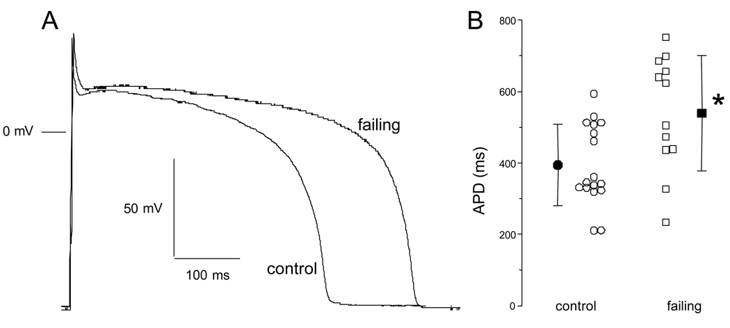
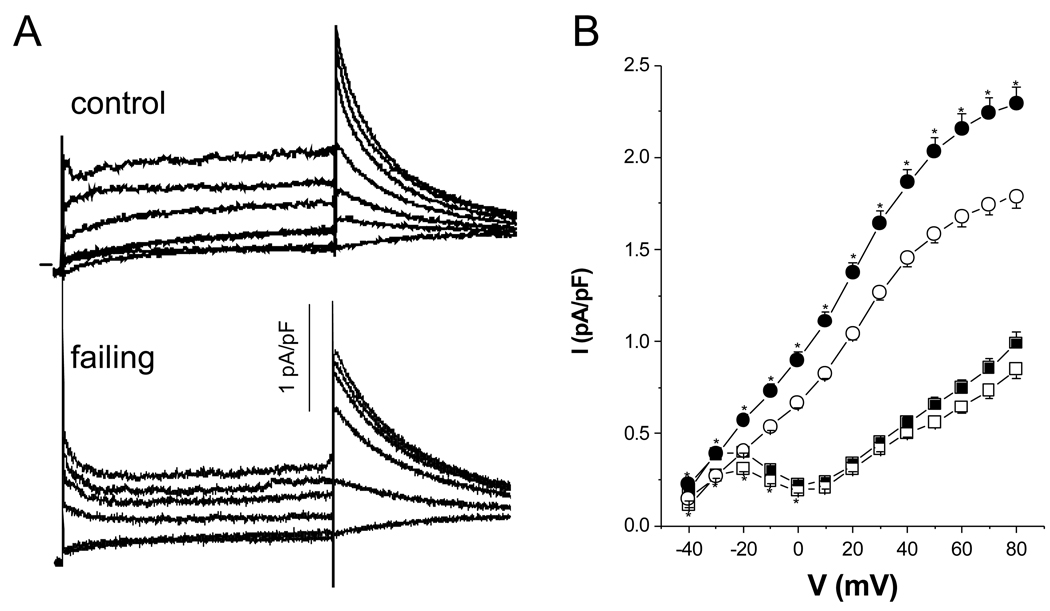
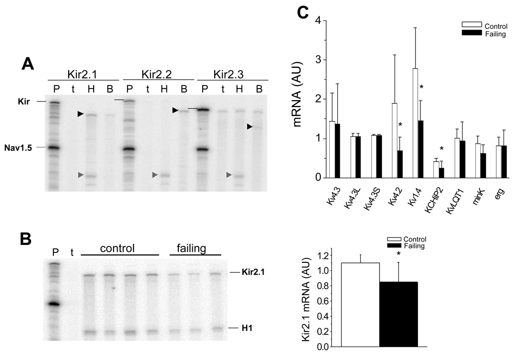
Action potential (AP) prolongation is a hallmark of failing myocardium. Functional downregulation of K currents is a prominent feature of cells isolated from failing ventricles. The detailed changes in K current expression differ depending on the species, the region of the heart, and the mechanism of induction of heart failure. We used complementary approaches to study K current downregulation in pacing tachycardia-induced heart failure in the rabbit. The AP duration (APD) at 90% repolarization was significantly longer in cells isolated from failing hearts compared with controls (539 +/- 162 failing vs. 394 +/- 114 control, P < 0.05). The major K currents in the rabbit heart, inward rectifier potassium current (I(K1)), transient outward (I(to)), and delayed rectifier current (I(K)) were functionally downregulated in cells isolated from failing ventricles. The mRNA levels of Kv4.2, Kv1.4, KChIP2, and Kir2.1 were significantly downregulated, whereas the Kv4.3, Erg, KvLQT1, and minK were unaltered in the failing ventricles compared with the control left ventricles. Significant downregulation in the long splice variant of Kv4.3, but not in the total Kv4.3, Kv4.2, and KChIP2 immunoreactive protein, was observed in cells isolated from the failing ventricle with no change in Kv1.4, KvLQT1, and in Kir2.1 immunoreactive protein levels. Multiple cellular and molecular mechanisms underlie the downregulation of K currents in the failing rabbit ventricle.


Figures

Figure 1
Figure 1
Differences in the action potential shape and duration in ventricular myocytes isolated from control and failing ventricles. A. The action potential profile in a cell isolated from a failing heart is characterized by a longer duration compared to a cell isolated from a control heart. B. Summarized data for action potential duration at 90% repolarization (APD90) in cells isolated from control (N=12 rabbits, n=17 cells) and failing (N=5 rabbits, n=12 cells) hearts. Despite a sizable variability, there is an overall prolongation of action potential duration in cells isolated from failing hearts. Mean±SD, *: p<0.05 vs. control.
Figure 2
Figure 2
Current density of inward rectifier current (IK1) in ventricular myocytes isolated from control and failing ventricles. Representative families of currents recorded from a holding potential of −20 mV in response to voltage steps of 500 ms from −150 mV to 50 mV in 20 mV increments in cells isolated from control (A) and failing (B) myocardium. The tracings shown are Ba2+-sensitive currents. The horizontal lines to the left of the current records indicate the zero-current level. C. The steady-state inward IK1 density is significantly reduced in cells isolated from failing (-■-, N=4 rabbits, n=8 cells) compared to cells isolated from control myocardium (-○-, N=4, n=9). D. The outward component of IK1 tends to decrease but the difference between cells isolated from control and failing hearts did not reach statistical significance. Mean ± SEM, *: p<0.05 vs. control.
Figure 3
Figure 3
Current density and kinetics of the calcium-independent transient outward current (Ito) in ventricular myocytes isolated from control and failing myocardium. Representative families of currents recorded from a holding potential of −80 mV in response to voltage steps of 500 ms from −40 mV to +80 mV in 20 mV increments in cells isolated from control (A) and failing (B) ventricles. The horizontal lines to the left of the current records indicate the zero-current level. C. The peak Ito density is significantly reduced in cells isolated from failing (-■-, N=11 rabbits, n=22 cells) compared to cells isolated from control myocardium (-○-, N=11, n=25). D. There was no difference in either the activation or the steady-state inactivation curves between cells isolated from control and failing myocardium. The fits to the data points are Boltzmann functions (control: -○-, N= 8, n=13, 27.3±1.8 mV half activation, 23.2±2.2 mV−1 maximal slope; N=6, n=7, −30.9±0.8 mV half inactivation, 9.9±0.7 mV−1 maximal slope; failing: -■-, N=7, n=10, 26.5±0.9 mV half activation, 19.1±1.0 mV−1 maximal slope; N=4, n=4, −25.3±1.0 mV half inactivation, 10.3±0.9 mV−1 maximal slope). E. There was no difference in the time constant of the macroscopic current relaxation (τ) between cells from control (-○-, N= 10, n= 20) and failing myocardium (-■-, N=5, n=11) determined by a single exponential fit of the first 150 ms of the current decay. F. Recovery from inactivation between cells from control (-○-, N=5, n=7, τ values of 120±45 ms and 1103±651 ms) and failing hearts (-■-, N=4, n=4, t values of 67±33 ms and 588±215 ms) did not differ. The lines were best fits to a biexponential function yielding the above-mentioned t. Mean ± SEM, *: p<0.05 vs. control.
Figure 4
Figure 4
A. Whole- cell IK currents recorded in cells isolated from control and failing ventricles.. The cells are held at −80 mV and step currents are elicited by voltage pulses from −50 to 80 mV in increments of 10 mV for 3 seconds. Tail currents are measured on return to −30 mV. For clarity every other current trace is shown. The currents were recorded with 10 mM KCl in the extracellular solution. B. The step current is significantly reduced at negative voltages in cells isolated from failing (-□-, N=5 rabbits, n=36 cells) compared to cells isolated from control myocardium (-■-, N=6, n=38). The tail current is significantly reduced in cells isolated from failing (-○-, N=5 rabbits, n=36 cells) compared to cells isolated from control myocardium over the entire voltage range (-•-, N=6, n=38). Mean±SEM, *: p<0.05 vs. control.
Figure 5
Figure 5
Relationship between action potential duration and Ito density in rabbit left ventricular myocytes isolated from the subendocardial and subepicardial layers of the left ventricle. A. Action potential and Ito recordings at +60 mV in cells from the subendocardial and subepicardial layers of the left ventricle at 37 °C. The myocyte from the subendocardial layer was characterized by a long action potential duration and a moderate Ito density compared to the cell isolated from the subepicardial layer. B. A plot of the correlation between Ito density at +60 mV and action potential duration at 90 % repolarization (APD90). There is a negative correlation between native Ito density and the APD90 (y = −14.8x + 410, n = 18, r = −0.80, p < 0.05), i.e. the higher the Ito density the shorter the action potential duration.
Figure 6
Figure 6
mRNA levels of K channel subunits in the failing heart. (A). RPAs for Kir2.x mRNAs in rabbit heart and brain. The horizontal black lines identify the positions of the Kir2.x probes and the arrowheads represent the Kir2.x protected fragments. The abbreviations are P: probes, t: yeast tRNA; H: heart; B: brain. Only Kir2.1 is detected in rabbit ventricle, Kir2.1, Kir2.2 and Kir2.3 transcripts are detected in rabbit brain. B. A representative RPA measuring Kir2.1 in 4 control and 3 failing rabbit ventricles. A bar-plot demonstrating that steady-state Kir2.1 mRNA normalized to Nav1.5 is decreased in failing compared to control ventricles (Nc=10; Nf=10, p = 0.009). C. Real-time PCR quantification of steady-state mRNA for Kv4.3, Kv4.3L, Kv4.3S, (Nc=8; Nf=7, p = NS), Kv4.2 (Nc=8; Nf=7, p = 0.027), Kv1.4 (Nc=8; Nf=7, p = 0.0096), KChIP2 (Nc=9, Nf=9; p = 0.028), KvLQT1 (Nc=9, Nf=7; p = NS), minK (Nc=8; Nf=6, p = 0.055), Erg (Nc=8; Nf=7, p = NS) and RPA for Kir2.1 (Nc=10; Nf=10, p = 0.0093) normalized to 28S mRNA in control and failing rabbit ventricles (AU: Arbitrary Units).
Figure 7
Figure 7
Representative Western blots together with summary data for K channel subunits. A. Protein levels of putative Ito subunits. Antibodies to Kv4.2 exhibit a single major band at 70 kDa which was significantly decreased in failing hearts (Nc=6; Nf=5, p < 0.05). Two different anti-Kv4.3 antibodies were used, an antibody that recognizes total Kv4.3 exhibits two major bands at 78 and 68 kDa that were quantified (Nc=6; Nf=6; p = NS). The primary antibody was specific for the long splice variant, anti-Kv4.3L, recognized a single major band at 78 kDa that was significantly reduced in failing ventricles (Nc=8; Nf=8; p = 0.0003). Anti-Kv1.4 antibodies recognize a single band at 96 kDa that was unchanged in the failing hearts (Nc=12; Nf=10, p = NS). Two anti-KChIP antibodies were used, an antibody specific for KChIP2S/T isoforms reveals two bands at 25 and 26 kDa that were unchanged in the failing heart (Nc=4; Nf=8, p = NS). A pan-KChIP antibody recognizes a single major band at 34 kDa that was significantly decreased in the failing myocardium (Nc=7; Nf=7, p < 0.05). B. Delayed rectifier immunoreactive protein. Single bands at ∼70 kDa were recognized with antibodies specific for KvLQT1 (Nc=10; Nf=11; p = NS), there was no significant change in failing hearts compared with controls. C. The anti-Kir2.1 antibody recognizes one band at 55 kDa that was unchanged in the failing hearts (Nc=6; Nf=8, p = NS). AU: Arbitrary Units.

References

    1. Ahmmed GU, Dong PH, Song G, Ball NA, Xu Y, Walsh RA, Chiamvimonvat N. Changes in Ca(2+) cycling proteins underlie cardiac action potential prolongation in a pressure-overloaded guinea pig model with cardiac hypertrophy and failure. Circ Res. 2000;86:558–570. - PubMed
    1. An WF, Bowlby MR, Betty M, Cao J, Ling HP, Mendoza G, Hinson JW, Mattsson KI, Strassle BW, Trimmer JS, Rhodes KJ. Modulation of A-type potassium channels by a family of calcium sensors. Nature. 2000;403:553–556. - PubMed
    1. Antzelevitch C, Sicouri S, Litovsky SH, Lukas A, Kishnan SC, Di Diego JM, Gintant GA, Liu D-W. Heterogeneity within the ventricular wall Electrophysiology and pharmacology of epicardial, endocardial, and M cells. Circ Res. 1991;69:1427–1449. - PubMed
    1. Armoundas AA, Wu R, Juang G, Marban E, Tomaselli GF. Electrical and structural remodeling of the failing ventricle. Pharmacol Ther. 2001;92:213–230. - PubMed
    1. Beuckelmann DJ, Nabauer M, Erdmann E. Alterations of K+ currents in isolated human ventricular myocytes from patients with terminal heart failure. Circ Res. 1993;73:379–385. - PubMed

Publication types

MeSH terms