. 2005 Apr 15;105(8):3222-9.
doi: 10.1182/blood-2004-11-4205. Epub 2005 Jan 6.

IgE- and IgE+Ag-mediated mast cell migration in an autocrine/paracrine fashion

Affiliations

IgE- and IgE+Ag-mediated mast cell migration in an autocrine/paracrine fashion

Jiro Kitaura et al. Blood. .

Abstract

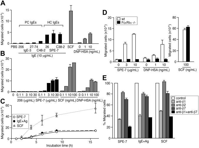
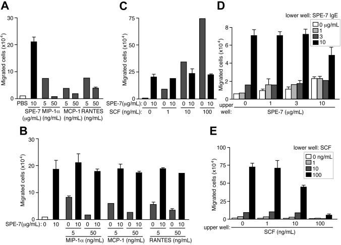
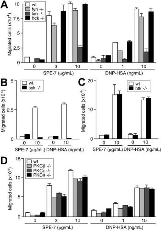
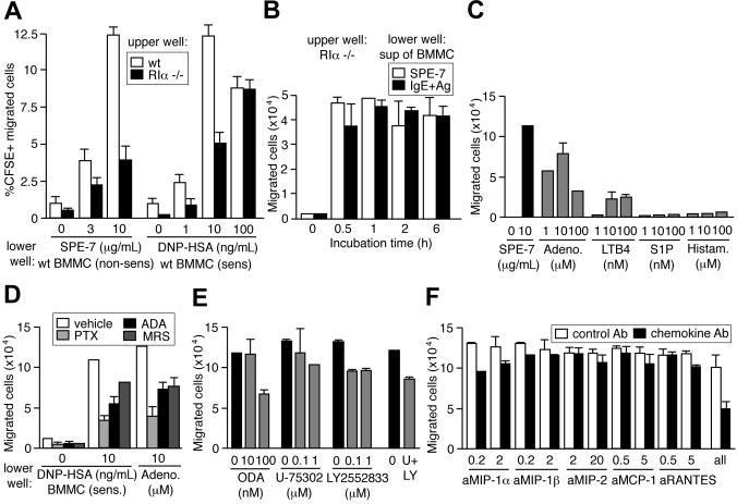
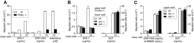
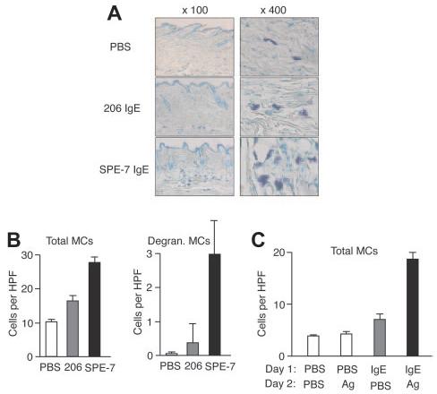
Mast cells are the major effector cells for immediate hypersensitivity and chronic allergic reactions. These cells accumulate in mucosal tissues of allergic reactions, where immunoglobulin E (IgE) is produced locally. Here we provide evidence that, in addition to antigen that can attract IgE-bound mast cells, the type of IgE molecules that efficiently activate mast cells can promote the migration of mast cells in the absence of antigen. IgE- and IgE+Ag-mediated migration involves an autocrine/paracrine secretion of soluble factors including adenosine, leukotriene B4, and several chemokines. Their secretion depends on 2 tyrosine kinases, Lyn and Syk, and they are agonists of G-protein-coupled receptors and signal through phosphatidylinositol 3-kinase gamma, leading to mast cell migration. In mouse experiments, naive mast cells are attracted to IgE, and IgE-sensitized mast cells are attracted to antigen. Therefore, IgE and antigen are implicated in mast cell accumulation at allergic tissue sites with local high IgE levels.


Figures

Figure 1
Figure 1
HC IgEs and IgE+Ag induce the migration of BMMCs through Fc∈;RI and β1 integrin. (A) HC, but not PC, IgEs induce BMMC migration efficiently in an 8-hour transwell assay. BMMCs, sensitized overnight with anti-DNP IgE (206) and washed, were also attracted to a lower well containing DNP-HSA. For comparison, SCF (100 ng/ml) was also tested in the same assay. Also shown are dose-response (B) and time-course (C) experiments using 10 μg/mL 206 IgE, 10 μg/mL SPE-7 IgE, 10 ng/mL DNP-HAS, and 100 ng/mL SCF. (D) Migration assays were performed with Fc∈;RIα/ and control BMMCs. (E) Migration of wild-type BMMCs was inhibited by neutralizing antibodies against integrins. Experiments were performed in triplicate. Representative data (± SD) are shown out of 2 or more experiments.
Figure 2
Figure 2
Comparison between HC IgEand chemokine-induced migration of BMMCs and checkerboard analysis of HC IgEinduced mast cell migration. (A) SPE-7 IgE–induced migration of BMMCs was compared with those induced by mast cell–attracting chemokines, MIP-1α, MCP-1, and RANTES. (B-C) BMMC migration was induced by SPE-7 IgE in combination with chemokines or SCF. (D) BMMCs in upper wells containing various concentrations of SPE-7 IgE were incubated with various concentrations of SPE-7 IgE in lower wells. (E) BMMCs in upper wells containing various concentrations of SCF were incubated with various concentrations of SCF in lower wells. Values (mean ± SD) are shown out of 2 independent experiments.
Figure 3
Figure 3
Effects of Src, Syk, Btk, and PKC deficiencies on IgE-induced migration. IgE- and IgE+Ag-induced migration assays were performed using Src family PTKdeficient (A), Syk-deficient (B), Btk-deficient (C), and PKC-deficient (D) BMMCs. Values (mean ± SD) are shown out of 2 independent experiments.
Figure 4
Figure 4
Involvement of adenosine, LTB4, and chemokines in an autocrine/paracrine mechanism of IgEand IgE+Ag-induced mast cell migration. (A) CFSE-labeled wild-type and Fc∈;RIα/ BMMCs in upper wells were attracted to lower wells containing wild-type cells and SPE-7 or 206 IgE–sensitized wild-type cells and antigen. (B) Supernatants of wild-type BMMCs incubated with SPE-7 for the indicated periods attracted Fc∈;RIα/ cells. Supernatants of PC IgE (206)–sensitized wild-type cells incubated with antigen for the indicated periods also attracted Fc∈;RIα/ cells. (C) Migration was induced by adenosine and LTB4, but not by sphingosine 1–phosphate (S1P) or histamine. (D) Pertussis toxin (PTX), adenosine deaminase (ADA), and adenosine A3 receptor inhibitor MRS 1523 inhibit the migration of CFSE-labeled wild-type BMMCs from upper wells to lower wells containing IgE-sensitized wild-type cells and antigen or adenosine. (E) 5-Lipoxygenase inhibitor (N-oleoyl dopamine [ODA]), BLT1 receptor inhibitor (U-75302), and BLT2 inhibitor (LY2552833) inhibit the migration of CFSE-labeled wild-type BMMCs from upper wells to lower wells containing IgE-sensitized wild-type cells and antigen. U-75302 and LY2552833 at 1 μM each were used in a combination (U+LY) as well. (F) Neutralizing antibodies to several chemokines inhibit the migration of CFSE-labeled wild-type BMMCs from upper wells to lower wells containing IgE-sensitized wild-type cells and antigen. All the antibodies at the higher concentrations each were used in a combination (all) as well. Mean values ± SD are shown out of 2 independent experiments.
Figure 5
Figure 5
IgE- and IgE+Ag-induced mast cell migration can be divided into the early phase of Lyn/Syk-dependent release of soluble factors and the later PI3Kγ-dependent phase. (A) PI3Kγ/ BMMCs were defective in migration in responses to SPE-7 IgE, IgE+Ag, or adenosine. (B) Fc∈;RIα/ cells in upper wells were incubated with lower wells containing wild-type, lyn/, or syk/ cells in the presence of SPE-7 or SCF. (C) Wild-type, lyn/, or syk/ cells in upper wells were incubated with lower wells containing 206 IgE–sensitized wild-type cells in the presence of DNP-HSA or SCF. Mean values ± SD are shown from 2 (A) or 3 (B,C) independent experiments.
Figure 6
Figure 6
In vivo models for IgE- and IgE+Ag-induced mast cell accumulation. (A) Gauze strips spotted with 10 μg/mL 206 or SPE-7 IgE or PBS were applied to the shaved back skin of naive NC/Nga mice for 24 hours. The mice were killed and skin samples were prepared to stain mast cells. Representative photomicrographs are shown. (B) Total and degranulating mast cell numbers per high-power field were counted. (C) NC/Nga mice were first treated with anti-DNP (206) IgE or PBS on the back skin, and 1 day later the same area was applied with gauze strips spotted with DNP-HSA or PBS for 24 hours. Total mast cell numbers per high-power field were counted. Data (mean values ± SD) are shown representative of 3 similar experiments.

References

    1. Metzger H. The receptor with high affinity for IgE. Immunol Rev. 1992;125:37–48. - PubMed
    1. Kinet JP. The high-affinity IgE receptor (Fc∈;RI): from physiology to pathology. Annu Rev Immunol. 1999;17:931–972. - PubMed
    1. Turner H, Kinet JP. Signalling through the high-affinity IgE receptor Fc∈;RI. Nature. 1999;402:B24–30. - PubMed
    1. Rivera J. Molecular adapters in Fc∈;RI signaling and the allergic response. Curr Opin Immunol. 2002;14:688–693. - PubMed
    1. Dastych J, Costa JJ, Thompson HL, Metcalfe DD. Mast cell adhesion to fibronectin. Immunology. 1991;73:478–484. - PMC - PubMed

Publication types