. 2005 Mar 1;386(Pt 2):221-6.
doi: 10.1042/BJ20041973.

Hepatically derived selenoprotein P is a key factor for kidney but not for brain selenium supply

Affiliations

Hepatically derived selenoprotein P is a key factor for kidney but not for brain selenium supply

Ulrich Schweizer et al. Biochem J. .

Abstract

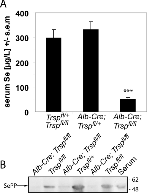
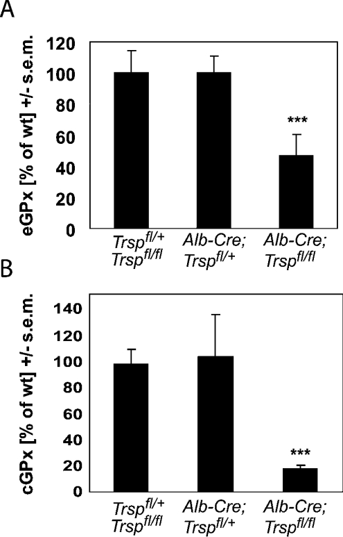
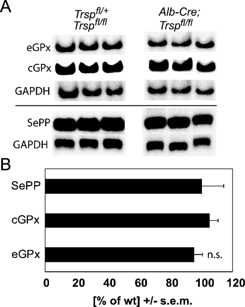
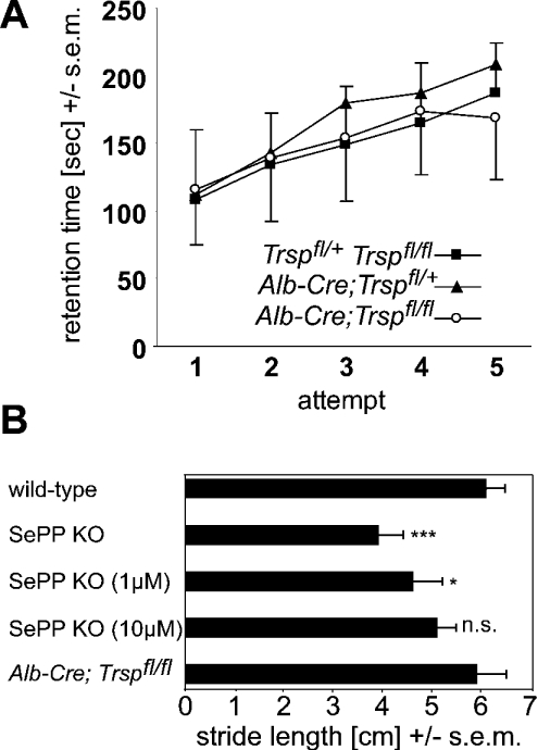
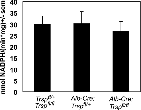
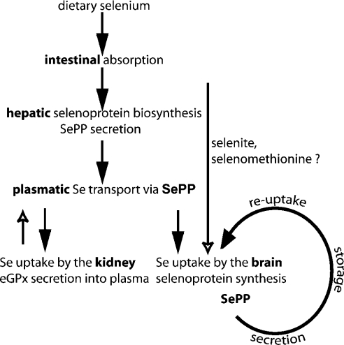
Liver-specific inactivation of Trsp, the gene for selenocysteine tRNA, removes SePP (selenoprotein P) from plasma, causing serum selenium levels to fall from 298 microg/l to 50 microg/l and kidney selenium to decrease to 36% of wild-type levels. Likewise, glutathione peroxidase activities decreased in plasma and kidney to 43% and 18% respectively of wild-type levels. This agrees nicely with data from SePP knockout mice, supporting a selenium transport role for hepatically expressed SePP. However, brain selenium levels remain unaffected and neurological defects do not occur in the liver-specific Trsp knockout mice, while SePP knockout mice suffer from neurological defects. This indicates that a transport function in plasma is exerted by hepatically derived SePP, while in brain SePP fulfils a second, hitherto unexpected, essential role.


Figures

Figure 1
Figure 1. Reduced SePP expression in liver-specific selenoprotein KO mice
(A) Serum Se levels in male mice at 5 weeks of age. The following genotypes were analysed: Trspfl/fl and Trspfl/+ (n=14), Alb-Cre; Trspfl/+ (n=10), and Alb-Cre; Trspfl/fl (n=8). Significant difference compared with wild type: ***P<0.001 (one-way ANOVA with Dunnett's post hoc test). (B) Western blot reveals a marked decrease in SePP expression in livers from liver-specific selenoprotein KO mice. Samples from individual mice of the indicated genotypes are shown. Serum served as a positive control for SePP. Positions of marker proteins in kDa are indicated.
Figure 2
Figure 2. Kidney selenoprotein expression depends on liver-derived SePP
(A) eGPx activity was measured in the plasma of 5-week-old male mice. The following genotypes were analysed: Trspfl/fl and Trspfl/+ (n=8), Alb-Cre; Trspfl/+ (n=10), and Alb-Cre; Trspfl/fl (n=7). (B) cGPx activity was determined in kidney homogenates with t-butylhydroperoxide as a substrate (male mice; n=6–9). Wild-type (Trspfl/fl and Trspfl/+) GPx activity was 516±60 nmol of NADPH/min per mg of protein. Significant differences compared with wild type: ***P<0.001 (one-way ANOVA with Dunnett's post hoc test).
Figure 3
Figure 3. Selenoprotein transcript levels are not altered in kidneys from liver-specific selenoprotein KO mice
(A) Northern blot analysis of eGPx, cGPx and SePP mRNAs. GAPDH was used as a standard. One representative Northern blot from two independent experiments is shown. Levels were measured in kidneys from 5-week-old male mice (n=5–6). (B) Quantification of selenoprotein mRNA expression. Phosphorimager signals were normalized using GAPDH. Results for liver-specific selenoprotein KO mice are expressed as a fraction of those for wild-type (wt) littermate controls. n.s., not significant (Student's t test).
Figure 4
Figure 4. Unlike SePP KO mice, liver-specific selenoprotein KO mice do not display a movement disorder
(A) Rotarod analysis. No differences in motor co-ordination were observed between genotypes Trspfl/fl and Trspfl/+ (n=21), Alb-Cre; Trspfl/+ (n=12), and Alb-Cre; Trspfl/fl (n=15). (B) Footprint analysis. Stride length is compared between SePP KO mice, SePP KO mice rescued with selenite from birth (1 μM or 10 μM in drinking water) and liver-specific selenoprotein KO mice. Selenite supplementation dose-dependently leads to amelioration of the movement disorder of SePP KO mice, while liver-specific selenoprotein KO mice are indistinguishable from wild-type mice. Results from wild-type littermate controls of each group were similar. (n=4–6). Significance of differences: n.s. not significant; *P<0.05; ***P<0.001 (one-way ANOVA with Dunnett's post hoc test).
Figure 5
Figure 5. Brain GPx activity is unaltered in liver-specific selenoprotein KO mice
GPx activity was determined in brain homogenates from 5-week-old mice with t-butylhydroperoxide as a substrate. The following genotypes were analysed: Trspfl/fl and Trspfl/+ (n=16), Alb-Cre; Trspfl/+ (n=12), and Alb-Cre; Trspfl/fl (n=16).
Figure 6
Figure 6. Summary of proposed function for SePP in Se transport and storage
The current model states that dietary Se compounds are taken up by the liver, where selenoproteins, including SePP, are synthesized. SePP is secreted into plasma and provides Se to the rest of the body. The kidney takes up liver-derived SePP from plasma for biosynthesis of selenoproteins, including eGPx. A similar scenario was expected for brain. However, in the absence of hepatic selenoprotein biosynthesis, plasma SePP and plasma Se levels are severely reduced, but brain selenoprotein metabolism remains unaffected, indicating an alternative route for Se uptake available to the brain. We propose SePP to also serve as a local Se storage and recycling protein. All available Se not needed to maintain selenoenzyme activities is incorporated into SePP to serve as an extracellular Se store. After proteolytic cleavage, the Se-rich C-terminus is taken up by the neuron and re-used for selenoprotein biosynthesis. Thus genetic inactivation of SePP compromises brain selenoprotein metabolism and Se retention, while liver-specific inactivation of SePP does not affect brain Se status, selenoprotein biosynthesis or Se retention.

Comment in

References

    1. Rayman M. P. The importance of selenium to human health. Lancet. 2000;356:233–241. - PubMed
    1. Kryukov G. V., Castellano S., Novoselov S. V., Lobanov A. V., Zehtab O., Guigo R., Gladyshev V. N. Characterization of mammalian selenoproteomes. Science. 2003;300:1439–1443. - PubMed
    1. Brigelius-Flohé R. Tissue-specific functions of individual glutathione peroxidases. Free Radical Biol. Med. 1999;27:951–965. - PubMed
    1. Arthur J. R. The glutathione peroxidases. Cell. Mol. Life Sci. 2000;57:1825–1835. - PMC - PubMed
    1. Rundlof A. K., Arner E. S. Regulation of the mammalian selenoprotein thioredoxin reductase 1 in relation to cellular phenotype, growth, and signaling events. Antioxid. Redox Signalling. 2004;6:41–52. - PubMed

Publication types