

Simultaneous recovery of extracellular and intracellular DNA suitable for molecular studies from marine sediments
- PMID: 15640168
- PMCID: PMC544275
- DOI: 10.1128/AEM.71.1.46-50.2005
Simultaneous recovery of extracellular and intracellular DNA suitable for molecular studies from marine sediments
Abstract
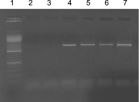
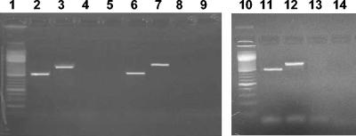
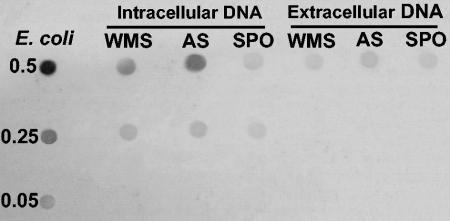
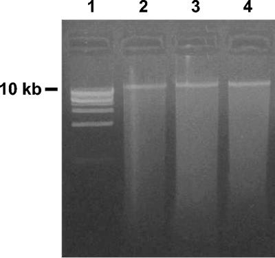
The occurrence of high extracellular DNA concentrations in aquatic sediments (concentrations that are 3 to 4 orders of magnitude greater than those in the water column) might play an important role in biogeochemical cycling, as well as in horizontal gene transfer through natural transformation. Since isolation of extracellular DNA from sediments is a difficult and unsolved task, in this study we developed an efficient procedure to recover simultaneously DNA associated with microbial cells and extracellular DNA from the same sediment sample. This procedure is specifically suitable for studying extracellular DNA because it avoids any contamination with DNA released by cell lysis during handling and extraction. Applying this procedure to different sediment types, we obtained extracellular DNA concentrations that were about 10 to 70 times higher than the intracellular DNA concentrations. Using specific targeted prokaryotic primers, we obtained evidence that extracellular DNA recovered from different sediments did not contain amplifiable 16S rRNA genes. By contrast, using DNA extracted from microbial cells as the template, we always amplified 16S rRNA genes. Although 16S rRNA genes were not detected in extracellular DNA, analyses of the sizes of extracellular DNA indicated the presence of high-molecular-weight fragments that might have contained other gene sequences. This protocol allows investigation of extracellular DNA and its possible participation in natural transformation processes.
Figures
References
-
- Atlas, R. M. 1993. Extraction of DNA from soils and sediments, p. 261-266 In P. F. Kemp, B. F. Sherr, and J. J. Cole (ed.), Handbook of methods in aquatic microbial ecology. Lewis Publishers, Boca Raton, Fla.
-
- Danovaro, R., A. Dell'Anno, A. Pusceddu, and M. Fabiano. 1999. Nucleic acid concentrations (DNA, RNA) in the continental and deep-sea sediments of the Eastern Mediterranean: relationships with seasonal varying organic inputs and bacterial dynamics. Deep-Sea Res. 46:1077-1094.
-
- DeFlaun, M. F., and W. H. Jeffrey. 1987. The distribution and molecular weight of dissolved DNA in subtropical estuarine and oceanic environments. Mar. Ecol. Prog. Ser. 38:65-73.
Publication types
MeSH terms
Substances
LinkOut - more resources
Full Text Sources
Other Literature Sources
