

Review
doi: 10.1186/gb-2004-6-1-202.
Epub 2004 Dec 21.
Profiling gene expression in growth-arrested mouse embryos in diapause
Affiliations
- PMID: 15642106
- PMCID: PMC549055
- DOI: 10.1186/gb-2004-6-1-202
Item in Clipboard
Review
Profiling gene expression in growth-arrested mouse embryos in diapause
Genome Biol.
2005.
Abstract
In many mammalian species, embryonic cell proliferation can be reversibly arrested in an embryonic diapause at the time of embryo implantation. A recent report has identified changes in embryonic gene expression that are associated with, and may halt, embryonic cell proliferation.
Figures

The growth of diapausing blastocysts is reversibly arrested before implantation into the uterus. (a) A diapausing blastocyst (arrowhead) is shown in contact with the uterine luminal epithelium (LE). Note the loose fibroblastic morphology of the stroma (S) underlying the luminal epithelium. T, the trophoblast of the blastocyst. (b) Implantation after diapause starts with the luminal epithelium adjacent to the trophoblast of the blastocyst undergoing apoptosis as the trophoblast cells start to invade the uterus. After implantation, the stroma has undergone massive proliferation and differentiation to form the decidua (D).
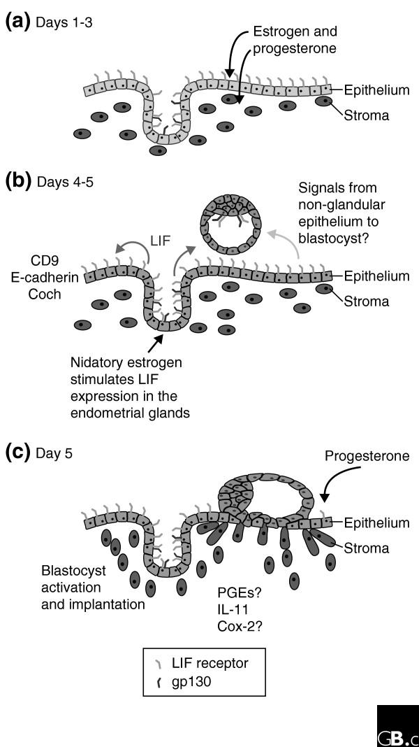
The time course of events leading to embryo implantation in the mouse. (a) On day 3 after fertilization the uterus is undergoing differentiation and proliferation under control of the ovarian steroid hormones estrogen and progesterone. (b) On day 4 the blastocyst is adjacent to the uterus and the production of leukemia inhibitory factor (LIF) is induced in the endometrial glands by nidatory estrogen. LIF is released into the uterine lumen, where it binds to LIF receptors on the luminal epithelium. LIF binding induces the expression of many genes, including the cell adhesion factors Coch and CD9, as the uterus becomes receptive to the embryo allowing the onset of implantation. Other adhesion molecules, such as E-cadherin, undergo redistribution in their expression. (c) On day 5 the blastocyst has started to invade the uterus and the stroma is undergoing decidualization, accompanied by the expression of prostaglandins (PGEs), which are regulated by Cox-2, and the cytokine interleukin-11 (IL-11), which is essential for decidualization.
Similar articles
-
Cloning of leukemia inhibitory factor (LIF) and its expression in the uterus during embryonic diapause and implantation in the mink (Mustela vison).Mol Reprod Dev. 1998 Sep;51(1):13-21. doi: 10.1002/(SICI)1098-2795(199809)51:1<13::AID-MRD2>3.0.CO;2-Z. Mol Reprod Dev. 1998. PMID: 9712313
-
Leukemia inhibitory factor can substitute for nidatory estrogen and is essential to inducing a receptive uterus for implantation but is not essential for subsequent embryogenesis.Endocrinology. 2000 Dec;141(12):4365-72. doi: 10.1210/endo.141.12.7855. Endocrinology. 2000. PMID: 11108244
-
Parthenogenic blastocysts cultured under in vivo conditions exhibit proliferation and differentiation expression genes similar to those of normal embryos.Anim Reprod Sci. 2011 Sep;127(3-4):222-8. doi: 10.1016/j.anireprosci.2011.08.005. Epub 2011 Aug 19. Anim Reprod Sci. 2011. PMID: 21890291
-
Leukaemia inhibitory factor in implantation and uterine biology.Reproduction. 2005 Aug;130(2):131-45. doi: 10.1530/rep.1.00304. Reproduction. 2005. PMID: 16049151 Review.
-
B1 and B2 Sox gene expression during neural plate development in chicken and mouse embryos: universal versus species-dependent features.Dev Growth Differ. 2011 Aug;53(6):761-71. doi: 10.1111/j.1440-169X.2011.01286.x. Epub 2011 Jul 18. Dev Growth Differ. 2011. PMID: 21762129 Review.
Cited by
-
Lactylation in cancer: Mechanisms in tumour biology and therapeutic potentials.Clin Transl Med. 2024 Nov;14(11):e70070. doi: 10.1002/ctm2.70070. Clin Transl Med. 2024. PMID: 39456119 Free PMC article. Review.
-
Gene expression, metabolic regulation and stress tolerance during diapause.Cell Mol Life Sci. 2010 Jul;67(14):2405-24. doi: 10.1007/s00018-010-0311-0. Epub 2010 Mar 7. Cell Mol Life Sci. 2010. PMID: 20213274 Free PMC article. Review.
-
An Integrative ATAC-Seq and RNA-Seq Analysis of the Endometrial Tissues of Meishan and Duroc Pigs.Int J Mol Sci. 2023 Sep 30;24(19):14812. doi: 10.3390/ijms241914812. Int J Mol Sci. 2023. PMID: 37834260 Free PMC article.
-
The molecular mechanisms of diapause and diapause-like reversible arrest.Biochem Soc Trans. 2023 Oct 31;51(5):1847-1856. doi: 10.1042/BST20221431. Biochem Soc Trans. 2023. PMID: 37800560 Free PMC article. Review.
-
The birth of embryonic pluripotency.Philos Trans R Soc Lond B Biol Sci. 2014 Dec 5;369(1657):20130541. doi: 10.1098/rstb.2013.0541. Philos Trans R Soc Lond B Biol Sci. 2014. PMID: 25349450 Free PMC article. Review.
References
-
- Stevens LC. The development of transplantable teratocarcinomas from intratesticular grafts of pre- and postimplantation mouse embryos. Dev Biol. 1970;21:364–382. - PubMed
-
- Psychoyos A. Endocrine control of egg implantation. In: Greep RO, Astwood EB, editor. In Handbook of Physiology. Vol. 2. Baltimore: Williams and Wilkins; 1973. pp. 187–215.
Publication types
MeSH terms
Substances
LinkOut - more resources
Full Text Sources
