. 2005;7(1):R46-59.
doi: 10.1186/bcr949. Epub 2004 Nov 8.

Breast fibroblasts modulate epithelial cell proliferation in three-dimensional in vitro co-culture

Affiliations

Breast fibroblasts modulate epithelial cell proliferation in three-dimensional in vitro co-culture

Andrea Sadlonova et al. Breast Cancer Res. 2005.

Abstract

Background: Stromal fibroblasts associated with in situ and invasive breast carcinoma differ phenotypically from fibroblasts associated with normal breast epithelium, and these alterations in carcinoma-associated fibroblasts (CAF) may promote breast carcinogenesis and cancer progression. A better understanding of the changes that occur in fibroblasts during carcinogenesis and their influence on epithelial cell growth and behavior could lead to novel strategies for the prevention and treatment of breast cancer. To this end, the effect of CAF and normal breast-associated fibroblasts (NAF) on the growth of epithelial cells representative of pre-neoplastic breast disease was assessed.

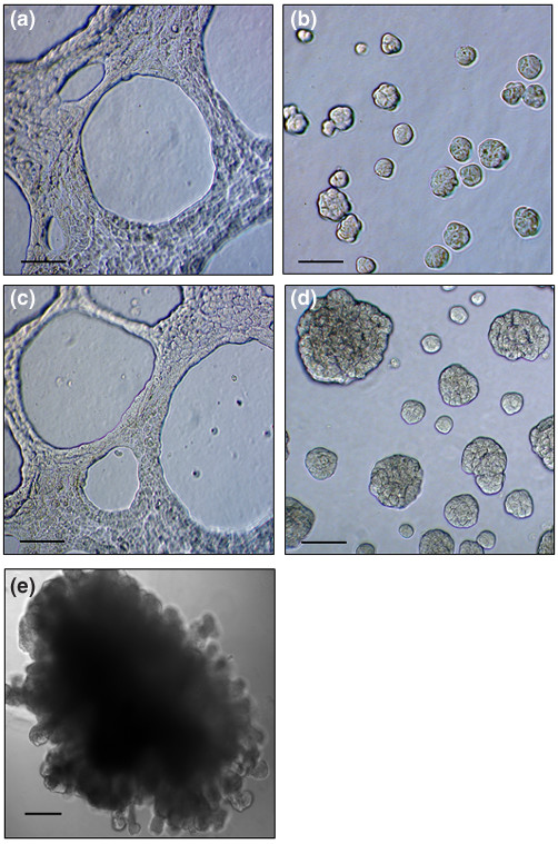
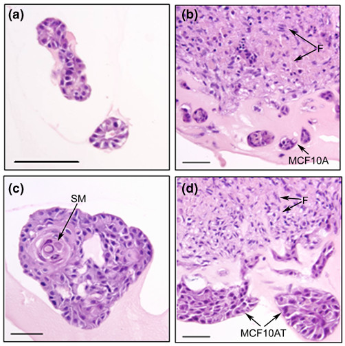
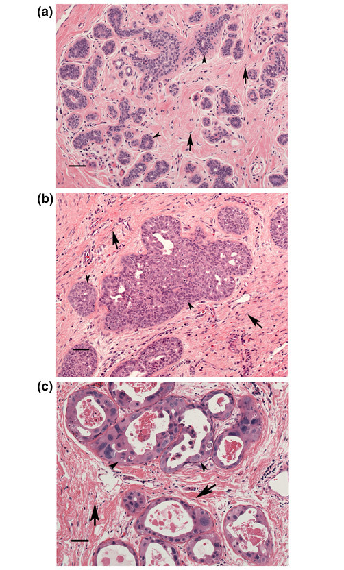
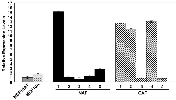
Methods: NAF and CAF were grown with the nontumorigenic MCF10A epithelial cells and their more transformed, tumorigenic derivative, MCF10AT cells, in direct three-dimensional co-cultures on basement membrane material. The proliferation and apoptosis of MCF10A cells and MCF10AT cells were assessed by 5-bromo-2'-deoxyuridine labeling and TUNEL assay, respectively. Additionally, NAF and CAF were compared for expression of insulin-like growth factor II as a potential mediator of their effects on epithelial cell growth, by ELISA and by quantitative, real-time PCR.

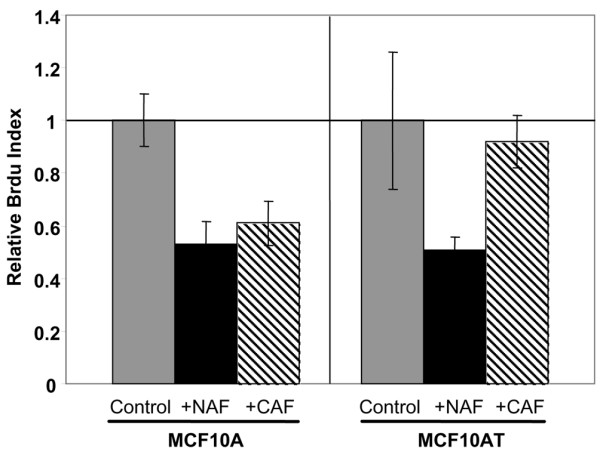
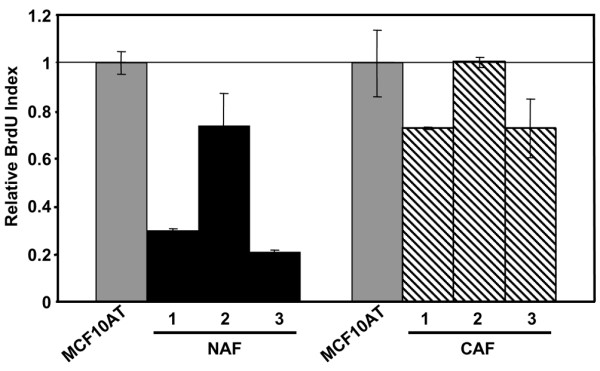
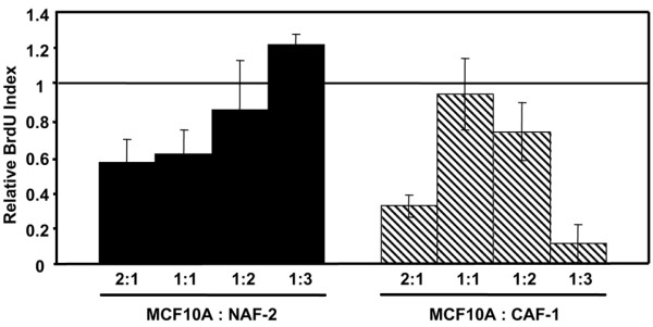
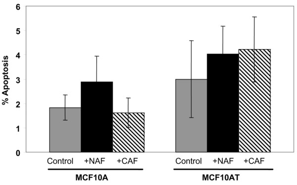
Results: In relatively low numbers, both NAF and CAF suppressed proliferation of MCF10A cells. However, only NAF and not CAF significantly inhibited proliferation of the more transformed MCF10AT cells. The degree of growth inhibition varied among NAF or CAF from different individuals. In greater numbers, NAF and CAF have less inhibitory effect on epithelial cell growth. The rate of epithelial cell apoptosis was not affected by NAF or CAF. Mean insulin-like growth factor II levels were not significantly different in NAF versus CAF and did not correlate with the fibroblast effect on epithelial cell proliferation.

Conclusion: Both NAF and CAF have the ability to inhibit the growth of pre-cancerous breast epithelial cells. NAF have greater inhibitory capacity than CAF, suggesting that the ability of fibroblasts to inhibit epithelial cell proliferation is lost during breast carcinogenesis. Furthermore, as the degree of transformation of the epithelial cells increased they became resistant to the growth-inhibitory effects of CAF. Insulin-like growth factor II could not be implicated as a contributor to this differential effect of NAF and CAF on epithelial cell growth.


Figures

Figure 1
Figure 1
MCF10A cells and MCF10AT cells in monoculture and in co-culture with fibroblasts. (a), (b) MCF10A cells and (c), (d) MCF10AT cells in monoculture initially form a lattice/scaffold arrangement (a, c). After several days of culture, spheroidal structures become more prominent (b, d). (e) MCF10AT cells in co-culture with fibroblasts form three-dimensional rounded structures. Similar structures are formed by MCF10A cells in co-culture with fibroblasts (phase contrast, 100 × magnification; scale bar, 200 μm).
Figure 2
Figure 2
H&E-stained histologic sections of MCF10A cell and MCF10AT cell monocultures and co-cultures with fibroblasts. (a) MCF10A cells form small spheroids. (b) MCF10A cells in co-culture with fibroblasts are located adjacent to the fibroblast aggregate (F) and maintain smaller spheroids. (c) In monoculture, MCF10AT cells form larger rounded three-dimensional structures. (d) In co-culture, MCF10AT cells form solid sheets and rounded groups of cells located adjacent to the fibroblasts (F). The occurrence of squamous metaplasia (SM) is more evident in MCF10AT monocultures, while it is suppressed in co-cultures. Overall, MCF10A cells have less squamous metaplasia than MCF10AT cultures (400 × magnification; scale bar, 50 μm).
Figure 3
Figure 3
Distribution and relative quantities of fibroblasts and epithelial cells in a normal terminal duct-lobular unit, hyperplasia and ductal carcinoma in situ (DCIS). (a) A terminal duct-lobular unit with epithelial cells (arrowheads) arranged in acini and intralobular terminal ducts separated by stroma-containing fibroblasts (arrows) in a ratio of epithelial cells to fibroblasts (E:F) of 2.7:1. (b) Ductal hyperplasia with a proliferation of epithelial cells (arrowheads) filling and expanding terminal ducts separated by reactive stroma including fibroblasts (arrows) in an E:F of 3.3:1. (c) High-grade DCIS, with epithelial cells (arrowheads) demonstrating markedly atypical nuclei, involving terminal ducts separated by reactive stroma including fibroblasts (arrows) in an E:F of 2:1 (200 × magnification; scale bar, 50 μm).
Figure 4
Figure 4
Proliferation of MCF10A cells and MCF10AT cells grown in monoculture and co-culture with fibroblasts. The rate of proliferation of MCF10A cells and MCF10AT cells, as measured by the 5-bromo-2'-deoxyuridine (BrdU) labeling index (assessed by immunocytochemistry), was significantly reduced in co-cultures of MCF10A cells with both normal breast-associated fibroblasts (NAF) (P = 0.009) and carcinoma-associated fibroblasts (CAF) (P = 0.024) compared with the MCF10A monoculture (control). The rate of proliferation of MCF10AT cells was significantly suppressed by NAF (P = 0.013) but not by CAF (P = 0. 935) in comparison with the MCF10AT monoculture (control).
Figure 5
Figure 5
5-Bromo-2'-deoxyuridine (BrdU) labeling, assessed by flow cytometry, of MCF10AT monocultures and co-cultures with normal breast-associated fibroblasts (NAF) and carcinoma-associated fibroblasts (CAF). These data are representative of replicate experiments indicating that NAF suppress proliferation of MCF10AT cells to a greater extent than do CAF. Again some variability in extent of suppression is present among individual NAF cultures and individual CAF cultures.
Figure 6
Figure 6
Relative 5-bromo-2'-deoxyuridine (BrdU) indices of MCF10A cells in co-culture with varying quantities of normal breast-associated fibroblast NAF-2 and carcinoma-associated fibroblast CAF-1. With increasing numbers of NAF-2, the mean rate of proliferation of co-cultured MCF10A cells increased, with a significant difference in BrdU-labeling index observed between a ratio of epithelial cells to fibroblasts (E:F) of 2:1 versus an E:F of 1:3 (P < 0.05). With increasing numbers of CAF-1, the mean rate of proliferation was highest at an E:F of 1:1. The rate of proliferation at an E:F of 1:1 was significantly higher than that at an E:F of 2:1 (P < 0.05). At an E:F of 1:3, CAF-1 caused a decreased proliferation of and enhanced cell death of MCF10A cells.
Figure 7
Figure 7
TUNEL assay for MCF10A cells and MCF10AT cells in co-culture with normal breast-associated fibroblasts (NAF) and carcinoma-associated fibroblasts (CAF). Co-culture with NAF and CAF had no significant effect on the rate of apoptosis of MCF10A cells and MCF10AT cells in comparison with epithelial cell monoculture controls.
Figure 8
Figure 8
Comparative expression of insulin-like growth factor (IGF) II mRNA. IGF II expression levels in MCF10A cells, MCF10AT cells, normal breast-associated fibroblasts (NAF) and carcinoma-associated fibroblasts (CAF) were determined by quantitative real-time PCR. All expression levels are relative to the calibrator, MCF10AT cells. The error bars represent the standard deviation of triplicate assays for each sample. Comparison of the mean expression level between NAF (mean = 4.2) and CAF (mean = 7.7) did not reach statistical significance (P = 0.39, t test).

References

    1. Kunz-Schughart LA, Knuechel R. Tumor-associated fibroblasts (part II): functional impact on tumor tissue. Histol Histopathol. 2002;17:623–637. - PubMed
    1. Kunz-Schughart LA, Knuechel R. Tumor-associated fibroblasts (part I): active stromal participants in tumor development and progression? Histol Histopathol. 2002;17:599–621. - PubMed
    1. Heffelfinger SC, Miller MA, Yassin R, Gear R. Angiogenic growth factors in preinvasive breast disease. Clin Cancer Res. 1999;5:2867–2876. - PubMed
    1. Costantini V, Sidoni A, Deveglia R, Cazzato OA, Bellezza G, Ferri I, Bucciarelli E, Nenci GG. Combined overexpression of urokinase, urokinase receptor, and plasminogen activator inhibitor-1 is associated with breast cancer progression: an immunohistochemical comparison of normal, benign, and malignant breast tissues. Cancer. 1996;77:1079–1088. doi: 10.1002/(SICI)1097-0142(19960315)77:6<1079::AID-CNCR12>3.0.CO;2-Z. - DOI - PubMed
    1. Brummer O, Athar S, Riethdorf L, Loning T, Herbst H. Matrix-metalloproteinases 1, 2, and 3 and their tissue inhibitors 1 and 2 in benign and malignant breast lesions: an in situ hybridization study. Virchows Arch. 1999;435:566–573. doi: 10.1007/s004280050442. - DOI - PubMed

Publication types