. 2005 Jan 17;168(2):303-13.
doi: 10.1083/jcb.200407072. Epub 2005 Jan 10.

Gp135/podocalyxin and NHERF-2 participate in the formation of a preapical domain during polarization of MDCK cells

Affiliations

Gp135/podocalyxin and NHERF-2 participate in the formation of a preapical domain during polarization of MDCK cells

Doris Meder et al. J Cell Biol. .

Abstract

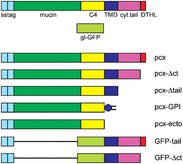
Epithelial polarization involves the segregation of apical and basolateral membrane domains, which are stabilized and maintained by tight junctions and membrane traffic. We report that unlike most apical and basolateral proteins in MDCK cells, which separate only after junctions have formed, the apical marker gp135 signifies an early level of polarized membrane organization established already in single cells. We identified gp135 as the dog orthologue of podocalyxin. With a series of domain mutants we show that the COOH-terminal PSD-95/Dlg/ZO-1 (PDZ)-binding motif is targeting podocalyxin to the free surface of single cells as well as to a subdomain of the terminally polarized apical membrane. This special localization of podocalyxin is shared by the cytoplasmic PDZ-protein Na+/H+ exchanger regulatory factor (NHERF)-2. Depleting podocalyxin by RNA interference caused defects in epithelial polarization. Together, our data suggest that podocalyxin and NHERF-2 function in epithelial polarization by contributing to an early apical scaffold based on PDZ domain-mediated interactions.


Figures

Figure 1.
Figure 1.
Gp135 marks a specialized apical domain in MDCK cells. PLAP-expressing MDCK cells were detached, fixed in suspension (A) or after plating onto coverslips (B, 1 h; C, 4 h), and stained with antibodies against apical (gp135, PLAP, and gp114) and basolateral (β1-integrin and E-cadherin) marker proteins. (A) Gp135 is distributed evenly over the whole surface of freshly detached and resuspended MDCK cells. (B) In single attached cells, gp135 is restricted to a preapical pole whereas other apical and basolateral marker proteins are not polarized. (C) Side views and confocal midsections of cell islands demonstrate that gp135 antibodies stain only the free surface, whereas PLAP, gp114, and E-cadherin are distributed randomly and localize to cell–cell contacts. The tight junction protein claudin-1 is still intracellular. Side views are merges of five vertical sections of confocal stacks. (D) Gp135 (red) is excluded from the center and the outer rim of the apical membrane of terminally polarized MDCK cells, whereas PLAP (green) is distributed over the entire apical membrane with only a small exclusion area in the center. Shown are confocal sections acquired from the same plane of the apical plasma membrane. (E) The exclusion area in the center of the apical membrane (gp135, red) corresponds to the site of outgrowth of the primary cilium as visualized by tubulin staining (green). Shown is a merged image of confocal sections taken of the apical membrane and the region above. Bars, 5 μm. (F) Overview of gp135 localization during the polarization of MDCK cells. Single, resuspended MDCK cells display an even distribution of plasma membrane proteins. Upon contact with a solid support, the cells establish a dynamic basal perimeter that includes apical membrane proteins (green) but not gp135 (red). When the cells flatten out and migrate toward each other, the basal plasma membrane and the edges of migrating cells are devoid of gp135. Upon cell–cell contact, gp135 disappears from the contacting membranes while other apical proteins remain. Apical-basolateral segregation starts at around 24 h and is completed after 3 d. In terminally polarized cells, gp135 is confined to a subdomain of the apical membrane that excludes the outer rim and the center surrounding the site of outgrowth of the primary cilium. Note that the membrane of the primary cilium (black) is devoid of classical apical marker proteins.
Figure 2.
Figure 2.
Purification of gp135. (A) Characterization of gp135 for enrichment from MDCK cells. Treatment with the nonionic detergent Triton X-100 and 1 mM EDTA on ice solubilized the majority of gp135 (S). Soluble and insoluble (IS) fractions were analyzed for their binding to wheat germ agglutinin (WGA). Shown is the analysis of the unbound fraction. All of gp135 is retained on the WGA column, whereas gp114 binds only partially. (B) Coomassie stained gel for identification of gp135. MDCK cells were treated with nonionic detergent on ice and solubilized glycoproteins enriched on a WGA column. Immunoprecipitation with mAbs against gp135 yielded a band at 135 kD. IgG hc corresponds to the heavy chain.
Figure 3.
Figure 3.
Overview of podocalyxin domain mutants. sstag, signal sequence followed by VSV-G epitope tag; mucin, Ser-Thr rich O-glycosylated domain; C4, tetracysteine stem region; TMD, transmembrane domain; cyt. tail, cytoplasmically oriented tail; DTHL, Asp-Thr-His-Leu COOH-terminal amino acids; gl-GFP, GFP containing two N-glycosylation sites; pcx, epitope tagged full-length podocalyxin; pcx-Δct, deletion of COOH-terminal DTHL motif; pcx-Δtail, deletion of cytoplasmic tail; pcx-GPI, GPI-anchored podocalyxin extracellular domain; pcx-ecto, soluble podocalyxin extracellular domain; GFP-tail, extracellular GFP with podocalyxin transmembrane domain and cytoplasmic tail; GFP-Δct, GFP-tail without COOH-terminal DTHL.
Figure 4.
Figure 4.
Localization of podocalyxin domain mutants. (A) Transiently transfected MDCK cells were analyzed 1 h after seeding. Epitope-tagged podocalyxin (pcx) colocalized to a high degree with endogenous podocalyxin, with both proteins being restricted to the free surface above the attachment area (top). In contrast, the domain mutant proteins often also localized to the dynamic basal perimeter of the cell (pcx-Δct; bottom). Endogenous podocalyxin is stained with the anti-gp135 antibody (red), and the mutant proteins are stained with anti-VSVG-tag antibody (green). Vertical sections of confocal stacks. Bars, 5 μm. (B) Scoring of localization patterns in single cells. “Like endogenous podocalyxin” means excluded from the basal perimeter. “Basal perimeter” refers to the additional presence in the dynamic basal extensions. Note that none of the mutant proteins showed efficient staining of the membrane in contact with the substratum. (C) MDCK cells 4 h after seeding. Confocal midsections reveal either staining of the free surface only (pcx; top) or localization to both the free surface and the contacting membrane (pcx-GPI, bottom). Bars, 5 μm. (D) Scoring of localization patterns in islands of cells. A phenotype similar to endogenous podocalyxin was scored as “only free surface.” Staining all over the plasma membrane was scored as “contacting membrane.” Pcx-GPI localizes to all plasma membrane domains. (E) Analysis of filter grown cells. Epitope-tagged podocalyxin (pcx) is excluded from the center region of the apical surface like endogenous podocalyxin. Pcx-Δct and pcx-GPI are also present on the ciliary membrane (insets). Note that pcx-GPI is even segregated from endogenous podocalyxin. Shown are confocal sections of the apical region of MDCK cells. The insets for pcx-Δct and pcx-GPI are taken from the central cell, 0.92 μm above the apical membrane. Bar, 10 μm. (F) Scoring of the restricted localization. A phenotype similar to endogenous podocalyxin is classified as “restricted,” and an unrestricted localization for the expressed mutant protein as “unrestricted.” The remaining percentage of transfected cells did not show an exclusion area for endogenous podocalyxin.
Figure 5.
Figure 5.
Apical sorting determinants within podocalyxin. (A) Polarized secretion of the soluble pcx-ectodomain. Terminally polarized, filter grown MDCK cells stably expressing pcx-ecto were analyzed by a secretion assay. Media supernatants were collected for 2 h. The apparent molecular mass of pcx-ecto is ∼110 kD. A, apical; B, basolateral. (B) Apical sorting of podocalyxin mutant proteins in stably expressing, terminally polarized cells. Depicted are epitope-tagged podocalyxin (pcx), GFP-tail, and GFP-Δct. GFP-tail lacking the extracellular domain of podocalyxin is still preferentially apical, whereas the corresponding protein without the four COOH-terminal amino acids appears unpolarized and partially intracellularly retained. Side views of polarized MDCK cell layers. Bar, 10 μm. (C) Quantification of apical and basolateral localization. Images were recorded so that the complete range of fluorescence intensities was included. Apical and basolateral membranes were outlined and fluorescence units calculated using Image J software. Relative apical and basolateral contributions were first calculated for individual cells and then for all cells of one population. Error bars are SD representing the variation of apical versus basolateral localization.
Figure 6.
Figure 6.
Podocalyxin knockdown by RNAi. (A) Knockdown efficiency for podocalyxin in MDCK cells. Total lysates (15 μg of protein) of knockdown cells (kd) were compared with lysates from control cells by Western blotting (ctrl; 1/1: 15 μg, 1/3: 5 μg, 1/10: 1.5 μg). Podocalyxin is reduced to ∼10% of wild-type levels in knockdown cells. Transferrin receptor (Trf-R) serves as a loading control. (B) Growth retardation of podocalyxin knockdown cells. 50,000 cells were seeded into individual 10-cm2 wells at day 0 and trypsinized cells counted each following day. Numbers of knockdown cells (kd) increased by an average of 1.7-fold per day in the first 4 d after seeding, whereas control cells (ctrl) multiplied at a rate of 3.2-fold per day. At 4 × 10⁶ cells/10 cm2 cells are confluent. (C) Immunofluorescence analysis of filter grown knockdown cells. Knockdown and control cells were seeded onto filters and fixed 2 d after reaching confluency. A confocal midsection stained for the basolateral marker gp58 shows that knockdown cells occupied a larger surface area. The corresponding vertical section shows that knockdown cells were less tall and did not restrict gp58 to the basolateral surface like control cells. The apical marker proteins gp114 and PLAP also displayed a less polarized distribution in knockdown cells. (D) Apical lumen formation in knockdown cells. Knockdown and control cells were suspended in Matrigel and cultured in the matrix for 4 d. Whereas >78% of the control cells had formed cysts with one central apical lumen as visualized by actin staining with Alexa Fluor 633-phalloidin and nuclear staining with DAPI, only 18% of knockdown cells displayed a single central lumen. 32% had no lumen and 50% displayed multiple small lumens. Bars, 10 μm.
Figure 7.
Figure 7.
Rescue of the polarization defect in knockdown cells by expression of GFP-podocalyxin. Knockdown cells, control cells, and knockdown cells stably expressing RNAi-resistant GFP-podocalyxin were seeded onto filters and fixed 2 d after reaching confluency. TRITC-phalloidin staining of actin shows that the GFP-pcx–expressing knockdown cells occupy a surface area similar to control cells and that they were almost as tall as control cells. Bars, 10 μm.
Figure 8.
Figure 8.
NHERF-2 shows a restricted localization similar to podocalyxin. MDCK cells stably expressing GFP-NHERF-2 were plated onto coverslips and fixed after 1 (A) and 4 h (B). (A) Side view of single cells. (B) Midsection through islands of cells viewed from the top. Both gp114 and β1-integrin show prominent staining of the contacting plasma membrane, whereas GFP-NHERF-2 and podocalyxin localize to the free surfaces surrounding the cell islands. Little GFP-NHERF-2 was observed at the cell–cell border. (C) Apical colocalization of GFP-NHERF-2 with podocalyxin in terminally polarized MDCK cells. Bars, 5 μm. (D) Interaction of podocalyxin and NHERF-2 at early stages of polarization. MDCK cells stably expressing GFP-NHERF-2 were allowed to attach for 1, 4, or 24 h. Postnuclear supernatants were immunoprecipitated with a GFP antibody. Western blotting with a podocalyxin antibody reveals increasing amounts from 1 to 24 h after seeding.

References

    1. Baas, A.F., J. Kuipers, N.N. van der Wel, E. Batlle, H.K. Koerten, P.J. Peters, and H.C. Clevers. 2004. Complete polarization of single intestinal epithelial cells upon activation of LKB1 by STRAD. Cell. 116:457–466. - PubMed
    1. Bacallao, R., C. Antony, C. Dotti, E. Karsenti, E.H. Stelzer, and K. Simons. 1989. The subcellular organization of Madin-Darby canine kidney cells during the formation of a polarized epithelium. J. Cell Biol. 109:2817–2832. - PMC - PubMed
    1. Balcarova-Stander, J., S.E. Pfeiffer, S.D. Fuller, and K. Simons. 1984. Development of cell surface polarity in the epithelial Madin-Darby canine kidney (MDCK) cell line. EMBO J. 3:2687–2694. - PMC - PubMed
    1. Bilder, D., M. Schober, and N. Perrimon. 2003. Integrated activity of PDZ protein complexes regulates epithelial polarity. Nat. Cell Biol. 5:53–58. - PubMed
    1. Bretscher, A., D. Chambers, R. Nguyen, and D. Reczek. 2000. ERM-Merlin and EBP50 protein families in plasma membrane organization and function. Annu. Rev. Cell Dev. Biol. 16:113–143. - PubMed

Publication types

MeSH terms