

Therapy of ocular and visceral leishmaniasis in a cat
- PMID: 15644104
- PMCID: PMC7169295
- DOI: 10.1111/j.1463-5224.2005.00342.x
Therapy of ocular and visceral leishmaniasis in a cat
Abstract
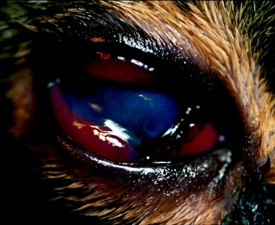
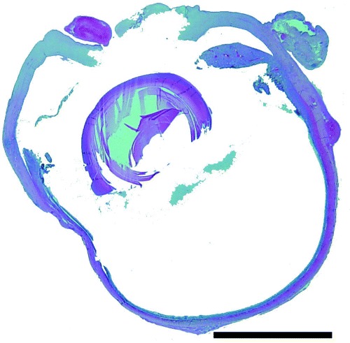
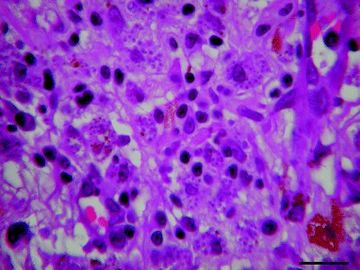
An 8-year-old, spayed female Domestic Short-haired cat was referred for further evaluation of chronic lymphocytic-plasmacytic stomatitis and bilateral ocular disease. The cat had been treated with systemic glucocorticoids for several months. Initial ophthalmic examination revealed bilateral deep stromal corneal ulcers, exudative panuveitis and secondary glaucoma. Mature mild neutrophilia and monocytosis were detected on complete blood cell count. Abnormalities in the serum profile were hyperglycemia, mild azotemia, hyperglobulinemia and moderate polyclonal gammapathy. Urinalysis revealed glucosuria without ketonuria. Diabetes mellitus was diagnosed and treatment with long-acting insulin was started. An enzyme-linked immunosorbent assay was highly positive for leishmaniasis, and treatment with allopurinol was started. Although specific topical treatment was applied, melting ulcers progressed to corneal perforation and both eyes were enucleated. Ocular histology showed large numbers of intracellular organisms compatible with amastigotes of the genus Leishmania located in the uveal tract, cornea, sclera and retina. Results of inmunohistochemistry staining on ocular samples were positive for Leishmania. Bone marrow cytology demonstrated numerous macrophages with intracytoplasmatic Leishmania. Polymerase chain reaction results on bone marrow for Leishmania were positive. Three weeks later, hypoglycemic episodes permitted withdrawal of the insulin therapy. To the authors' knowledge this is the first case of ocular and visceral leishmaniasis diagnosed in vivo and under systemic treatment in a cat.
Figures

References
-
- Slappendel RJ, Ferrer L. Leishmaniasis In: Microbiology and Infectious Diseases of Dogs and Cats. (ed. Green C.) W.B. Saunders, Philadelphia, 1998;. 450–458.
-
- Del Giudice P, Marty P. Cat‐associated zoonosis: don't forget rabies and leishmaniasis. Archives of Internal Medicine 2003; 62: 1945–1952. - PubMed
-
- Barnes JC, Stanley O, Craig TM. Diffuse cutaneous leishmaniasis in a cat. Journal of the American Veterinary Medical Association 1993; 202: 416–418. - PubMed
-
- Ozon C, Marty P, Pratlong F et al. Disseminated feline leishmaniosis due to Leishmania infantum in southern France. Veterinary Parasitology 1998;. 75: 273–277. - PubMed
Publication types
MeSH terms
Substances
LinkOut - more resources
Full Text Sources
Medical
Miscellaneous
