. 2005 Jan 18;102(3):725-30.
doi: 10.1073/pnas.0406509102. Epub 2005 Jan 11.

Syncytin-A and syncytin-B, two fusogenic placenta-specific murine envelope genes of retroviral origin conserved in Muridae

Affiliations

Syncytin-A and syncytin-B, two fusogenic placenta-specific murine envelope genes of retroviral origin conserved in Muridae

Anne Dupressoir et al. Proc Natl Acad Sci U S A. .

Abstract

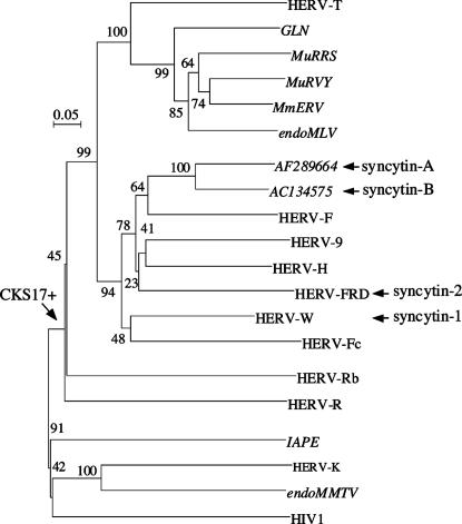
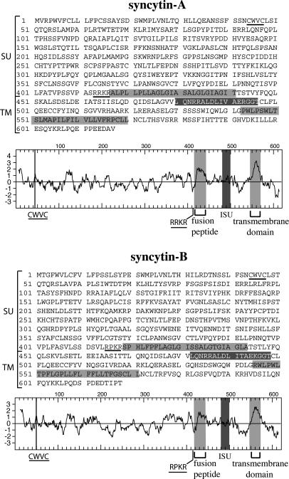
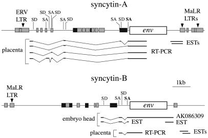
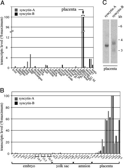
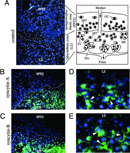
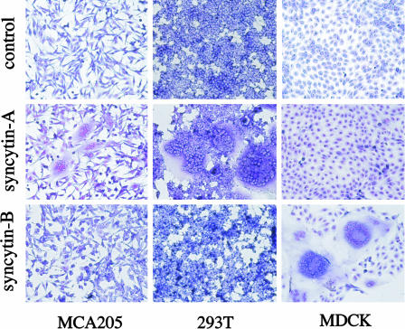
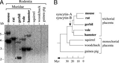
Recently, we and others have identified two human endogenous retroviruses that entered the primate lineage 25-40 million years ago and that encode highly fusogenic retroviral envelope proteins (syncytin-1 and -2), possibly involved in the formation of the placenta syncytiotrophoblast layer generated by trophoblast cell fusion at the materno-fetal interface. A systematic in silico search throughout mouse genome databases presently identifies two fully coding envelope genes, present as unique copies and unrelated to any known murine endogenous retrovirus, that we named syncytin-A and -B. Quantitative RT-PCR demonstrates placenta-specific expression for both genes, with increasing transcript levels in this organ from 9.5 to 14.5 days postcoitum. In situ hybridization of placenta cryosections further localizes these transcripts in the syncytiotrophoblast-containing labyrinthine zona. Consistently, we show that both genes can trigger cell-cell fusion in ex vivo transfection assays, with distinct cell type specificities suggesting different receptor usage. Genes orthologous to syncytin-A and -B and disclosing a striking conservation of their coding status are found in all Muridae tested (mouse, rat, gerbil, vole, and hamster), dating their entry into the rodent lineage approximately 20 million years ago. Together, these data strongly argue for a critical role of syncytin-A and -B in murine syncytiotrophoblast formation, thus unraveling a rather unique situation where two pairs of endogenous retroviruses, independently acquired by the primate and rodent lineages, would have been positively selected for a convergent physiological role.


Figures

Fig. 1.
Fig. 1.
Envelope-based phylogenetic tree with positions of syncytin-A and -B. The tree was determined by the neighbor-joining method by using TM envelope sequences (see ref. 17), essentially from murine ERVs (in italics) and human HERVs. The horizontal branch length and scale indicate the percentage of amino acid substitutions from the node, and the arrow on the left points to the node of the CKS17-positive sequence group. Percent bootstrap values obtained from 1,000 replicates are indicated. Accession numbers for each representative element together with the sequence alignment used to generate the tree are given in Fig. 8, which is published as supporting information on the PNAS web site.
Fig. 2.
Fig. 2.
Primary sequence, hydrophobicity profile, and predicted features of the syncytin-A and -B envelopes. The SU and TM moieties of the envelopes are delineated, with the canonical furin cleavage site (R/K-N-R/K-R) between the two subunits and the CWVC domain involved in SU–TM interaction underlined; the hydrophobic fusion peptide and TM domains are shaded in light gray, and the putative immunosuppressive domain (ISU) in dark gray.
Fig. 3.
Fig. 3.
Genomic organization of the syncytin-A and -B chromosomal locus. The envelope ORF (open box), the pol-related sequences (dark gray boxes), and murine repetitive elements (light-gray boxes) are indicated. Partial structure of envelope transcripts, as determined experimentally by RT-PCR (with reverse primers in the env ORF) or by an in silico search throughout EST and cDNA databases, are schematized, together with the related splice donor (SD) and splice acceptor (SA) sites positioned on the DNA sequences (with the canonical retroviral envelope SA site in bold).
Fig. 4.
Fig. 4.
Real-time quantitative RT-PCR and Northern blot analysis of syncytin-A and -B transcripts in mouse tissues. (A and B) syncytin-A (gray bars) and -B (dark bars) transcript levels in tissues of adult male (same results for the corresponding female tissues, when tested, namely thyroid, cervical node, pancreas, adrenal, salivary, and parotid glands; data not shown), adult female (uterus and ovaries), and 11.5-day-pregnant female (embryos and placenta) mice (A), and embryonic tissues (embryo, yolk sac, amnion, and placenta) at different times postcoitum (from day 7.5 to 16.5) (B). The embryo head (h) and body (b) were analyzed separately whenever possible. (C) Northern blot analysis of 11.5 dpc mouse placenta total RNA, probed with a syncytin-A or -B full-length env probe. Exposure time, 4 days.
Fig. 5.
Fig. 5.
In situ hybridization of 14.5 dpc mouse placenta for syncytin-A and -B expression. Scheme of a mouse mature placenta cross section: the labyrinthine zona (LZ) is the site of exchange between the maternal blood (Mb) and the fetal blood vessels (Fbv) and contains mononuclear trophoblast cells (MT) that terminally differentiate, by cell fusion, into syncytiotrophoblasts (ST) directly lining the fetal blood vessels; in Muridae, both cell types are arranged as a three-layered barrier (two syncytial and one mononuclear layer, not represented) between the maternal and fetal blood circulation; the spongiotrophoblast zona with the spongiotrophoblast cells (SPT) and the outermost layer with the trophoblast giant cells (GC) (producing hormone/growth factors) overlie the labyrinthine zona. (A) Negative control: tissue section probed with a digoxigenin-labeled sense syncytin-A riboprobe. (B and C) Tissue sections probed with digoxigenin-labeled antisense syncytin-A and -B riboprobes, respectively, disclosing hybridization signals only within the labyrinthine zona (the dotted lines delineate the separation between the labyrinthine and spongiotrophoblastic zona). (D and E) Magnification of the labyrinthine zona in B and C, with the arrowheads pointing to labeled structures with several nuclei. Nuclei were stained in blue by using DAPI. A–C, ×100; D and E, ×400.
Fig. 6.
Fig. 6.
Syncytin-mediated cell–cell fusion. Shown is syncytia formation by the syncytin-A and -B envelope genes. The indicated cells were transfected with expression vectors for syncytin-A or -B, or a negative control (syncytin-A in antisense orientation) and were stained with May–Grünwald and Giemsa 24–48 h after transfection.
Fig. 7.
Fig. 7.
Conservation of the syncytin-A and -B genes during rodent evolution. (A) Southern blot of DraI-restricted genomic DNA from the indicated rodent species, with the syncytin-B fragments (asterisk) and the syncytin-A fragments (arrowhead) both found in Muridae only (the syncytin-A bands have a lower intensity due to the use of a syncytin-B probe, with the opposite result when using a syncytin-A probe; not shown). (B) Schematized rodent phylogenetic tree, with the arrow indicating the date of entry of the two syncytin-A and -B envelope genes; the placenta type, in terms of structure of the syncytiotrophoblast materno–fetal barrier, is indicated (see Discussion).

References

    1. Boeke, J. D. & Stoye, J. P. (1997) in Retroviruses, eds. Coffin, J. M., Hughes, S. H. & Varmus, H. E. (Cold Spring Harbor Lab. Press, Plainview, NY), pp. 343-435. - PubMed
    1. Bannert, N. & Kurth, R. (2004) Proc. Natl. Acad. Sci. USA 13, 14572-14579. - PMC - PubMed
    1. de Parseval, N. & Heidmann, T., Cytogenet. Genome Res., in press. - PubMed
    1. Lander, E. S., Linton, L. M., Birren, B., Nusbaum, C., Zody, M. C., Baldwin, J., Devon, K., Dewar, K., Doyle, M., FitzHugh, W., et al. (2001) Nature 409, 860-921. - PubMed
    1. Waterston, R. H., Lindblad-Toh, K., Birney, E., Rogers, J., Abril, J. F., Agarwal, P., Agarwala, R., Ainscough, R., Alexandersson, M., An, P., et al. (2002) Nature 420, 520-562. - PubMed

Publication types