. 2005 Jan;144(1):17-27.
doi: 10.1038/sj.bjp.0706030.

Antimony-induced cardiomyopathy in guinea-pig and protection by L-carnitine

Affiliations

Antimony-induced cardiomyopathy in guinea-pig and protection by L-carnitine

Marco Alvarez et al. Br J Pharmacol. 2005 Jan.

Abstract

Antimony (Sb) is the mainstay for the treatment of Leishmaniasis. It has serious, often lethal, cardiovascular side effects. The objective of this study was to examine the effects of Sb treatment upon the electrocardiogram (ECG), myocyte contractility (assessed by monitoring sarcomere length during field stimulation), whole-cell action potential (AP) and calcium current (I(Ca)) of the guinea-pig and to evaluate L-carnitine as a cardioprotective agent. Guinea-pigs received daily injections of either saline, Sb(V), Sb(III), L-carnitine or L-carnitine with Sb(III). Eight lead ECGs were recorded under halothane anaesthesia every 4 days. At the end of each treatment regime, animals were killed and ventricular myocytes were enzymatically isolated. Treatment with Sb(V) for 26 days prolonged the QT interval of the ECG. Treatment with Sb(III) was lethal within 2 days for approximately 50% of the animals. The survivors showed ECG alterations similar to those described in man: T wave flattening and/or inversion, depression of the ST segment, and elongation of RR and QT intervals. Their ventricular myocytes showed impaired contraction responses to changes in stimulus frequency, elongated AP and reduced I(Ca). Combined treatment with L-carnitine and Sb(III) delayed mortality. Prior treatment with L-carnitine followed by combined treatment with L-carnitine and Sb(III) reduced mortality to <10% over 12 days and these animals showed normal ECG. Their myocytes showed normal contractility and AP. It is concluded that L-carnitine has a preventive cardioprotective role against antimony-induced cardiomyopathy. The mechanism of action of L-carnitine may be to counter oxidative stress caused by Sb(III).


Figures

Figure 1
Figure 1
Typical eight lead electrocardiogram from a guinea-pig. The three classical frontal leads D1, D2 and D3 were recorded simultaneously. The three augmented leads aVR, aVL and aVF were recorded consecutively. The two precordial leads V1 and V6 were recorded simultaneously. Time and voltage scale apply to all traces. Dotted lines represent 0 mV.
Figure 2
Figure 2
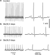
Effects of Sb(III) on the guinea-pig ECG. ECGs were recorded from one animal before (a) and 4 days (b) and 8 days (c) following the onset of daily injections of Sb(III). All traces represent the D2 lead of the ECG, details of which are shown upon an expanded time scale to the right. Time scales apply to traces in the respective columns. The vertical scale applies to all traces. The dotted lines indicate 0 mV.
Figure 3
Figure 3
Effects of Sb(III) on wave and interval durations (a) and wave amplitudes (b) of the guinea-pig ECG. Measurements were performed before and following 4 days and 8 days of Sb(III) injections. All measurements were performed on the D2 lead. (a) RR, PQ, QRS and QT are the classical intervals, QRS being measured as a QJ interval. Classical QT, measured from the beginning of Q to the end of T, is presented with its corrected values using either Bazett formula (QTcb) or Fridericia formula (QTcf). Pdur: duration of the P waves. JT+: interval from the J point to the maximum of the T wave. T+Te: interval from the maximum to the end of the T wave. (b) In addition to the classical waves (P, Q, R, S and T), J point and ST elevation are also shown. Columns and bars represent mean±s.e. (n=13). Significance according to paired Student's t-test between control and Sb(III) treatments are indicated as * (P<0.05) or ** (P<0.001).
Figure 4
Figure 4
Cartesian representation of the guinea-pig ECG and its modification by Sb(III). Coupled derivations are in (A), D2 versus D1 and in (B), V1 versus V6. Data were recorded from the same animal before (a) and following 8 days (b) treatment with Sb(III). Each trace contains about 25 cycles (loops) corresponding to 30 s acquisition.
Figure 5
Figure 5
AP and calcium currents recorded from enzymatically dissociated ventricular myocytes. (a) Superimposed typical AP recorded in myocytes from three different guinea-pigs treated with either saline, L-carnitine or Sb(III) for 8 days. (b, c) Typical calcium currents recorded in normal Tyrode solution, in myocytes from the same guinea-pigs as in (a), with a voltage step to 0 mV following a 100 ms prepulse from −80 to −40 mV: saline (b) or Sb(III) (c)-treated guinea-pigs. The cell's capacitances were 37 and 35 pF for the saline and Sb(III) myocytes, respectively. Time and amplitude scales apply to both (b, c) panels. The zero current level is indicated by the horizontal line near each current trace. The stimulation frequency was 0.2 Hz in (a), and 0.1 Hz in (b, c).
Figure 6
Figure 6
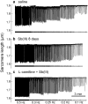
Effect of Sb(III) upon stimulus frequency-dependent changes of SL in isolated ventricular myocytes of guinea-pig. SL was measured by an on-line video detection method (see Methods) where downward deflection of the trace represents sarcomere shortening upon field stimulation which is schematically indicated below each recording. (a) A myocyte obtained from a control animal injected daily with saline. (b) A myocyte obtained from an animal injected daily with Sb(III) for 8 days. (c) A myocyte obtained from an animal injected with L-carnitine alone for 4 days followed by injection with L-carnitine and Sb(III) for 12 days. The time scale in (c) is applicable to all three traces. In each cell, stimulus frequency was progressively reduced from 0.5 Hz to 0.3 Hz, 0.25 Hz, 0.2 Hz and 0.1 Hz as indicated below the recordings.
Figure 7
Figure 7
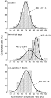
The effect of Sb(III) upon the normalised contraction–amplitude ratio of isolated venticular myocytes from guinea-pig. Contraction–amplitude ratio represents sarcomere shortening recorded at 0.5 Hz over sarcomere shortening recorded at 1 Hz. Results are shown for myocytes from control animals injected with saline (a; N=9 guinea-pigs; n=75 cells), myocytes from guinea-pigs injected with Sb(III) for 8 days (b; N=8; n=76) and myocytes from guinea-pigs treated for 4 days with L-carnitine and then for 12 days with L-carnitine plus Sb(III) (c; N=7; n=70). The lines represent gaussian functions fit to the data with their associated mean and s.d. values. Distributions were tested for normality with the Anderson–Darling normality test. In myocytes from Sb(III)-injected animals, the distribution needs two gaussians. A contraction–amplitude ratio of 100% indicates no change in amplitude with frequency at 0.5 and 1 Hz.
Figure 8
Figure 8
Contractions of isolated ventricular myocytes of guinea-pig. Typical contractions elicited by field stimulation (squares) from myocytes isolated from a guinea-pig injected with (a) saline, (b) Sb(III) for 8 days and (c) 4 days with L-carnitine and then for 12 days with L-carnitine plus Sb(III). Each set in (a, b and c) represents superimposed contractions obtained at stimulation frequencies of 0.3 and 1 Hz. Horizontal bar (200 ms) and SL scale apply to all three sets. (d–f) Multiphasic contractions obtained under similar conditions from myocytes isolated from 8 days Sb(III)-treated guinea-pigs. Contractions induced by field stimulation (squares) are followed by after-contractions (arrows). Contractions illustrated in (b and d) are from the same guinea-pig.

References

    1. BALSHAW D.M., XU L., YAMAGUCHI N., PASEK D.A., MEISSNER G. Calmodulin binding and inhibition of cardiac muscle calcium release channel (ryanodine receptor) J. Biol. Chem. 2001;276:20144–20153. - PubMed
    1. BERESEWICZ A., HORACKOVA M. Alterations in electrical and contractile behavior of isolated cardiomyocytes by hydrogen peroxide: possible ionic mechanisms. J. Mol. Cell. Cardiol. 1991;23:899–918. - PubMed
    1. BERS D.M. Excitation–Contraction Coupling and Cardiac Contractile Force 2001Boston: Kluwer Academic Press Publishers; 2nd edn.
    1. BORASO A., WILLIAMS A.J. Modification of the gating of the cardiac sarcoplasmic reticulum Ca(2+)-release channel by H2O2 and dithiothreitol. Am. J. Physiol. 1994;267:H1010–H1016. - PubMed
    1. BRADLEY W.R., FREDERICK W.G. The toxicity of antimony. Ind. Med. 1941;2:15–22.

Publication types

MeSH terms