. 2005 Mar;76(3):510-6.
doi: 10.1086/428141. Epub 2005 Jan 11.

Identification of C7orf11 (TTDN1) gene mutations and genetic heterogeneity in nonphotosensitive trichothiodystrophy

Affiliations

Identification of C7orf11 (TTDN1) gene mutations and genetic heterogeneity in nonphotosensitive trichothiodystrophy

Kazuhiko Nakabayashi et al. Am J Hum Genet. 2005 Mar.

Abstract

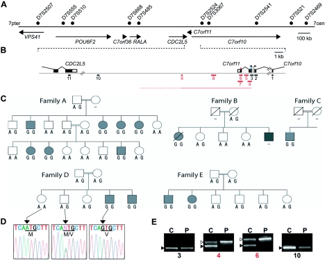
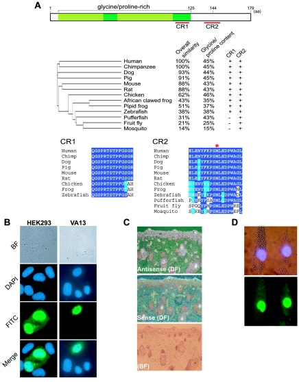
We have identified C7orf11, which localizes to the nucleus and is expressed in fetal hair follicles, as the first disease gene for nonphotosensitive trichothiodystrophy (TTD). C7orf11 maps to chromosome 7p14, and the disease locus has been designated "TTDN1" (TTD nonphotosensitive 1). Mutations were found in patients with Amish brittle-hair syndrome and in other nonphotosensititive TTD cases with mental retardation and decreased fertility but not in patients with Sabinas syndrome or Pollitt syndrome. Therefore, genetic heterogeneity in nonphotosensitive TTD is a feature similar to that observed in photosensitive TTD, which is caused by mutations in transcription factor II H (TFIIH) subunit genes. Comparative immunofluorescence analysis, however, suggests that C7orf11 does not influence TFIIH directly. Given the absence of cutaneous photosensitivity in the patients with C7orf11 mutations, together with the protein's nuclear localization, C7orf11 may be involved in transcription but not DNA repair.


Figures

Figure  1
Figure 1
Identification of C7orf11 mutations. To screen the two exons and the 5′ upstream region of the C7orf11 gene, we used three sets of primer pairs: C7orf11-5upF/ex1R1, C7orf11ex1-F2/R3, and C7orf11ex2-F/R2 (for primer sequences, see table A1 [online only]). The cycling conditions were initial denaturation at 94°C for 3 min followed by 35 cycles of denaturation at 94°C for 30 s, annealing at 58°C for 30 s, and extension at 72°C for 60 s. PCR products were purified using microCLEAN (Microsone) and were used as the sequencing template. Sequencing reactions were performed using the Big Dye Terminator kit (Applied Biosystems), and an ABI-3730 DNA Sequencer (Applied Biosystems) was used to obtain sequences. A, Physical map of the 2-Mb region on 7p14 showing homozygosity in a consanguineous Amish kindred. B, Diagram of C7orf11, consisting of two exons spanning 2 kb. The coding and untranslated regions are shown as blackened and unblackened boxes, respectively. The red and blue asterisks (*) indicate the position of the M144V mutation in the Amish kindred and the 2-bp deletion in the Moroccan siblings with TTD, respectively. Black and red bars (numbered from 1 to 11) represent the size and location of the PCR products in the deletion mapping for patient 6474. The fragments that were not amplified from patient 6474 (because of homozygous deletion) are indicated by red bars. C, Five affected families from the Amish kindred. Gray and blackened symbols indicated affected members, and unblackened symbols indicate unaffected members. The member of family B represented by the blackened symbol is the proband, who had a more severe phenotype, presumably due to a de novo chromosomal abnormality (46,XY,14q) (Jackson et al. 1974). The genotype at the M144V mutation site (A, wild-type; G, mutated) is shown for each member. D, Electropherograms for the M144V site. E, Representative results (from test primers 3, 4, 6, and 10) of the PCR-based deletion mapping of the C7orf11 locus in patient 6474 (lane C, control; lane P, patient 6474). Unblackened triangles indicate the 704-bp fragment amplified by control primer DJg5/g6. The blackened triangles indicate the fragment amplified by a test primer pair. The cycling conditions were initial denaturation at 94°C for 3 min followed by 36 cycles of denaturation at 94°C for 30 s, annealing at 58°C for 30 s, and extension at 72°C for 75 s. Detailed information for the 11 test primers pairs and the control primer pair is available in table A1 (online only).
Figure  2
Figure 2
A, The human C7orf11 protein. The glycine/proline–rich region is shown in green (the low-complexity regions detected by the BLASTP program are in light green). There are two highly conserved C terminal regions (CR1 and CR2) among the candidate orthologues. The evolutionary tree (middle) was drawn by the ClustalX program (Thompson et al. 1997). The protein sequences used for phylogenetic analysis are human C7orf11 (NCBI accession number NP_619646) and orthologues from chimpanzee (Ensembl accession number ENSPTRP00000032652), mouse (NCBI accession number BAB27916), rat (Ensembl accession number ENSRNOP00000018746), chicken (Ensembl accession number ENSGALP00000020100), frog (Xenopus tropicalis [NCBI accession number NP_989025]), pufferfish (Tetraodon nigroviridis [NCBI accession number CAF91712]), fruit fly (Drosophila melanogaster [NCBI accession number NP_648690]), and mosquito (Anopheles gambiae [NCBI accession number XP_318005]). Deduced protein sequences were derived from cDNA sequences from zebrafish (Danio rerio [GenBank accession number BC062385]) and pig (GenBank accession number BP456435). The dog protein sequence was predicted from the genomic DNA sequence (see UCSC Genome Browser Web site). For each species, the overall amino acid similarity with the human protein sequence, the percentage ratio of glycine/proline content in the region from the N terminus to CR1, and the presence (+) or absence (−) of CR1 and CR2 are shown. Multiple sequence alignments for CR1 and CR2 are at bottom. The position of M144V in CR2 is indicated by a red asterisk (*). Identical and similar amino acids are highlighted in dark and light blue, respectively. B, Subcellular localization assay. Transiently expressed Myc-tagged C7orf11 in human cultured cells (transformed human embryonic kidney cells [HEK293] and SV40-transformed WI38 human embryonic fibroblasts [VA13]) was examined by immunostaining, by use of anti-Myc antibody and a secondary antibody conjugated with fluorescein isothiocyanate (FITC). Bright-field (BF), 4'-6-diamidino-2-phenylindole (DAPI), FITC, and the merged image of DAPI and FITC signals are shown for each cell line. The Myc-tagged cDNA construct for C7orf11 was generated by inserting the coding region of C7orf11 (nucleotides 51–590 of mRNA) in pcDNA3.1+ Myc-His A (Invitrogen). DNA transfection was performed using Lipofectamine Plus (Invitrogen). Two days after transfection, cells were washed in PBS and were fixed in 50% acetone plus 50% methanol at −20°C for 15 min. The samples were blocked with 10% BSA in PBS (blocking buffer) for 1 h and were incubated in blocking buffer containing anti-Myc antibody (Santa Cruz Biotechnology) for 45 min. Cells were washed three times with PBS, were incubated with FITC-conjugated anti-mouse IgG antibody for 30 min, and were washed three times with PBS. After DAPI staining, the samples were analyzed by deconvolution microscopy (Zeiss). C, In situ RNA hybridization for C7orf11 on human fetal skin tissue. A 556-bp cDNA fragment (corresponding to nucleotides 303–858 of mRNA) was subcloned in pCR-Script (Stratagene) and was used as template DNA to synthesize [35S]UTP-labeled riboprobes. [35S]UTP-labeled riboprobes (sense and antisense) were hybridized to the sections. Radioactive signals were detected by autoradiography with 38 d of exposure. Slides were counterstained with hematoxylin-eosin and were photographed under dark-field (DF) and bright-field (BF) illumination. The signal detected in epidermis and hair follicles is specific to the antisense probe. The detail conditions used for hybridization, washing, and signal detection were described elsewhere (Saarialho-Kere et al. 1992). D, Normal level of TFIIH in fibroblast cells from one of the Moroccan siblings with the 2-bp deletion. Mixed DAPI/phase contrast image (top) showing a normal (with large beads) and TTD (with small beads) cells. Immunofluorescence with anti-XPB antibody (bottom). For exact quantitative comparison of fluorescence signals between normal and TTD fibroblasts without slide-to-slide variability, two types of fibroblasts were preloaded with cytoplasmic plastic beads of different sizes (large beads for normal and small beads for TTD) and then were mixed, cultured overnight, and processed for immunofluorescence.

References

Electronic-Database Information

    1. The Chromosome 7 Annotation Project, http://www.chr7.org/ (for seven candidate genes on 7p14)
    1. Ensembl, http://www.ensembl.org/ (for proteins fromchimpanzee [accession number ENSPTRP00000032652],rat [accession number ENSRNOP00000018746], and chicken [accession number ENSGALP00000020100])
    1. GenBank, http://www.ncbi.nlm.nih.gov/Genbank/ (forhuman C7orf11 mRNA [accession number NM_138701] and cDNA sequences from zebrafish [accession number BC062385] and pig [accession number BP456435])
    1. NCBI, http://www.ncbi.nlm.nih.gov/ (for human C7orf11 [accession number NP_619646] and orthologues from mouse [accession number BAB27916], frog [accession number NP_989025], pufferfish [accession number CAF91712], fruitfly [accession number NP_648690], and mosquito [accession number XP_318005])
    1. Online Mendelian Inheritance in Man (OMIM), http://www.ncbi.nlm.nih.gov/Omim/ (for Amish brittle-hair syndrome, Sabinas syndrome, and Pollitt syndrome)

References

    1. Alfandari S, Delaporte E, van Neste D, Lucidarme-Delespierre E, Piette F, Bergoend H (1993) A new case of isolated trichothiodystrophy. Dermatology 186:197–200 - PubMed
    1. Baden HP, Jackson CE, Weiss L, Jimbow K, Lee L, Kubilus J, Gold RJ (1976) The physicochemical properties of hair in the BIDS syndrome. Am J Hum Genet 28:514–521 - PMC - PubMed
    1. Bergmann E, Egly JM (2001) Trichothiodystrophy, a transcription syndrome. Trends Genet 17:279–28610.1016/S0168-9525(01)02280-6 - DOI - PubMed
    1. Botta E, Nardo T, Broughton BC, Marinoni S, Lehmann AR, Stefanini M (1998) Analysis of mutations in the XPD gene in Italian patients with trichothiodystrophy: site of mutation correlates with repair deficiency, but gene dosage appears to determine clinical severity. Am J Hum Genet 63:1036–1048 - PMC - PubMed
    1. Botta E, Nardo T, Lehmann AR, Egly JM, Pedrini AM, Stefanini M (2002) Reduced level of the repair/transcription factor TFIIH in trichothiodystrophy. Hum Mol Genet 11:2919–292810.1093/hmg/11.23.2919 - DOI - PubMed

Publication types