

Non-traumatic cerebrospinal fluid rhinorrhea: diagnosis and management
- PMID: 15646164
- PMCID: PMC6147856
- DOI: 10.5144/0256-4947.2004.453
Non-traumatic cerebrospinal fluid rhinorrhea: diagnosis and management
Abstract
Background: Although the majority of cerebrospinal (CSF) fistulas in the anterior skull base are traumatic in nature, the minority is non-traumatic or primary. Non-traumatic CSF leak can be a diagnostic and treatment challenge.
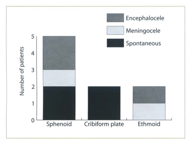
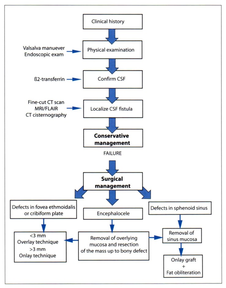
Patients and methods: We describe the diagnosis, modified methods of localization, and surgical repair of a series of nine patients who presented with non-traumatic CSF rhinorrhea and were managed between July 2000 and October 2002.
Results: Eight patients were managed via an endoscopic approach and one patient through an intracranial approach. The RI/T2-FLAIR test was used for localization of the site of the leak. The test confirmed the site of CSF leak in 6 patients. Successful repair of CSF rhinorrhea was achieved in 7 of 8 patients with a single endoscopic procedure; one patient required two procedures after a re-leak 18 months following the first repair.
Conclusion: Non-traumatic CSF rhinorrhea is a relatively rare condition and occurs secondary to different etiologies. Among multiple techniques available for localization, MRI/FLAIR is effective, but requires further evaluation and polishing. In the absence of a large skull base lesion or tumor, endoscopic repair of CSF fistula carries a high success rate with a high margin of safety and low morbidity rate.
Figures
Comment in
-
RE: Non-traumatic cerebrospinal fluid rhinorrhea: diagnosis and management.Ann Saudi Med. 2005 Nov-Dec;25(6):515. doi: 10.5144/0256-4947.2005.515. Ann Saudi Med. 2005. PMID: 16438469 Free PMC article. No abstract available.
Similar articles
-
Endoscopic management of cerebrospinal fluid rhinorrhea.Laryngoscope. 2004 Oct;114(10):1833-7. doi: 10.1097/00005537-200410000-00029. Laryngoscope. 2004. PMID: 15454781
-
Endoscopic CSF leak repair.Curr Opin Otolaryngol Head Neck Surg. 2007 Feb;15(1):35-9. doi: 10.1097/MOO.0b013e3280123fce. Curr Opin Otolaryngol Head Neck Surg. 2007. PMID: 17211181 Review.
-
Endoscopic repair of traumatic CSF rhinorrhea in a pediatric patient.Int J Pediatr Otorhinolaryngol. 1996 Jul;36(2):109-15. doi: 10.1016/0165-5876(96)01342-0. Int J Pediatr Otorhinolaryngol. 1996. PMID: 8818757
-
Endonasal endoscopic repair of cerebrospinal fluid rhinorrhea.Minim Invasive Neurosurg. 2004 Jun;47(3):173-7. doi: 10.1055/s-2004-818451. Minim Invasive Neurosurg. 2004. PMID: 15343435
-
Management of post-traumatic cerebrospinal fluid (CSF) leak of anterior skull base: 10 years experience.Acta Otolaryngol. 2013 Sep;133(9):944-50. doi: 10.3109/00016489.2013.793821. Acta Otolaryngol. 2013. PMID: 23944946 Review.
Cited by
-
RE: Non-traumatic cerebrospinal fluid rhinorrhea: diagnosis and management.Ann Saudi Med. 2005 Nov-Dec;25(6):515. doi: 10.5144/0256-4947.2005.515. Ann Saudi Med. 2005. PMID: 16438469 Free PMC article. No abstract available.
-
Multiple uses of fibrin sealant for nervous system treatment following injury and disease.J Venom Anim Toxins Incl Trop Dis. 2017 Mar 14;23:13. doi: 10.1186/s40409-017-0103-1. eCollection 2017. J Venom Anim Toxins Incl Trop Dis. 2017. PMID: 28293254 Free PMC article. Review.
-
Systematic Review: Success Rate of Endoscopic Endonasal versus Combined Endonasal and Transorbital Neuroendoscopic Approach for Nontraumatic Cerebrospinal Fluid Leak Repairs in the Lateral Recess of Sphenoid Sinus.J Neurol Surg B Skull Base. 2024 Apr 8;86(2):138-159. doi: 10.1055/s-0044-1785486. eCollection 2025 Apr. J Neurol Surg B Skull Base. 2024. PMID: 40104536 Free PMC article.
-
Endoscopic management of cerebrospinal fluid rhinorrhea.Asian J Neurosurg. 2016 Jul-Sep;11(3):183-93. doi: 10.4103/1793-5482.145101. Asian J Neurosurg. 2016. PMID: 27366243 Free PMC article. Review.
-
Diagnosis and Localization of Cerebrospinal Fluid Rhinorrhea: A Systematic Review.Am J Rhinol Allergy. 2022 May;36(3):397-406. doi: 10.1177/19458924211060918. Epub 2021 Nov 30. Am J Rhinol Allergy. 2022. PMID: 34846218 Free PMC article.
References
-
- Zapalac JS, Marple BF, Schwade ND. Skull base cerebrospinal fluid fistulas: A comprehensive diagnostic algorithm. Otolaryngol Head Neck Surg. 2002;126:669–676. - PubMed
-
- Dandy WD. Pneumocephalus (intracranial pneumocele or aerocele) Arch Surg. 1926;12:949–982.
-
- Park JI, Strezlow VV, Freidman WH. Current management of cerebrospinal fluid rhinorrhea. Laryngoscope. 1983;93:1294–1300. - PubMed
-
- Dohlman G. Spontaneous cerebrospinal rhinorrhea. Acta Otolaryngol Suppl (Stockh) 1948;67:20–23. - PubMed
MeSH terms
LinkOut - more resources
Full Text Sources