

Compartmental oxidation of thiol-disulphide redox couples during epidermal growth factor signalling
- PMID: 15647005
- PMCID: PMC1134784
- DOI: 10.1042/BJ20041829
Compartmental oxidation of thiol-disulphide redox couples during epidermal growth factor signalling
Abstract
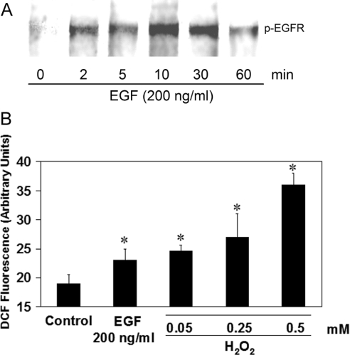
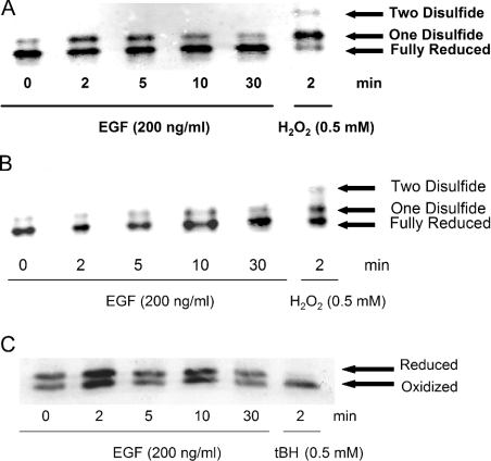
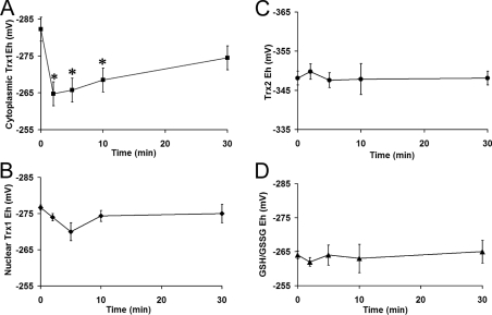
Exogenously added ROS (reactive oxygen species) cause generalized oxidation of cellular components, whereas endogenously generated ROS induced by physiological stimuli activate discrete signal transduction pathways. Compartmentation is an important aspect of such pathways, but little is known about its role in redox signalling. We measured the redox states of cytosolic and nuclear Trx1 (thioredoxin-1) and mitochondrial Trx2 (thioredoxin-2) using redox Western blot methodologies during endogenous ROS production induced by EGF (epidermal growth factor) signalling. The glutathione redox state was measured by HPLC. Results showed that only cytosolic Trx1 undergoes significant oxidation. Thus EGF signalling involves subcellular compartmental oxidation of Trx1 in the absence of a generalized cellular oxidation.
Figures
References
-
- Finkel T. Oxidant signals and oxidative stress. Curr. Opin. Cell Biol. 2003;15:247–254. - PubMed
-
- Droge W. Free radicals in the physiological control of cell function. Physiol. Rev. 2002;82:47–95. - PubMed
-
- Carpenter G., Cohen S. Epidermal growth factor. J. Biol. Chem. 1990;265:7709–7712. - PubMed
-
- Sundaresan M., Yu Z. X., Ferrans V. J., Irani K., Finkel T. Requirement for generation of H2O2 for platelet-derived growth factor signal transduction. Science. 1995;270:296–299. - PubMed
-
- Meng T. C., Fukada T., Tonks N. K. Reversible oxidation and inactivation of protein tyrosine phosphatases in vivo. Mol. Cell. 2002;9:387–399. - PubMed
Publication types
MeSH terms
Substances
Grants and funding
LinkOut - more resources
Full Text Sources
Miscellaneous