. 2005 Jan 12;4(1):2.
doi: 10.1186/1476-4598-4-2.

Stable expression of constitutively-activated STAT3 in benign prostatic epithelial cells changes their phenotype to that resembling malignant cells

Affiliations

Stable expression of constitutively-activated STAT3 in benign prostatic epithelial cells changes their phenotype to that resembling malignant cells

Hosea F Huang et al. Mol Cancer. .

Abstract

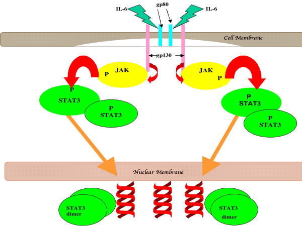
Background: Signal transducers and activators of transcription (STATs) are involved in growth regulation of cells. They are usually activated by phosphorylation at specific tyrosine residues. In neoplastic cells, constitutive activation of STATs accompanies growth dysregulation and resistance to apoptosis through changes in gene expression, such as enhanced anti-apoptotic gene expression or reduced pro-apoptotic gene expression. Activated STAT3 is thought to play an important role in prostate cancer (PCA) progression. Because we are interested in how persistently-activated STAT3 changes the cellular phenotype to a malignant one in prostate cancer, we used expression vectors containing a gene for constitutively-activated STAT3, called S3c, into NRP-152 rat and BPH-1 human benign prostatic epithelial cells.

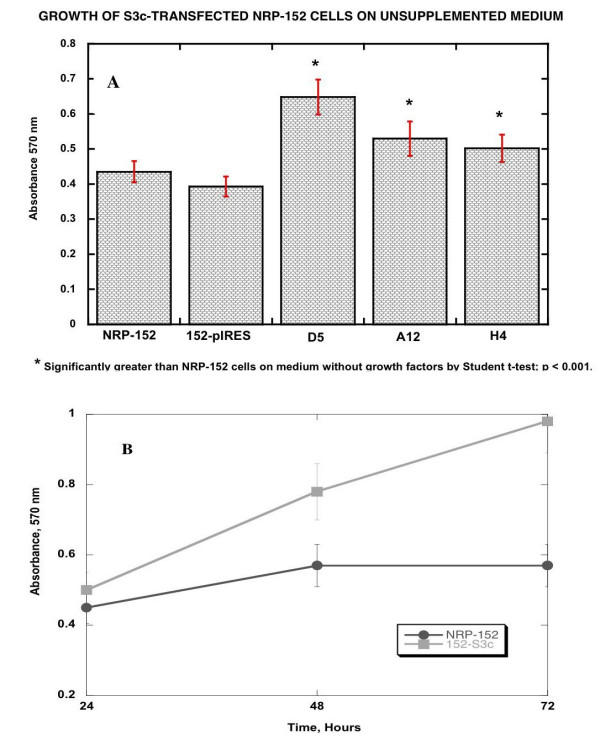
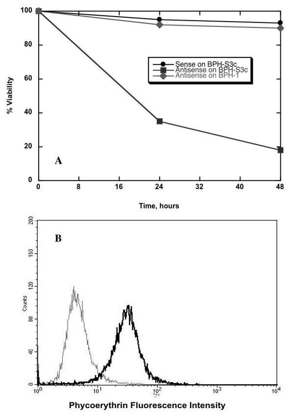
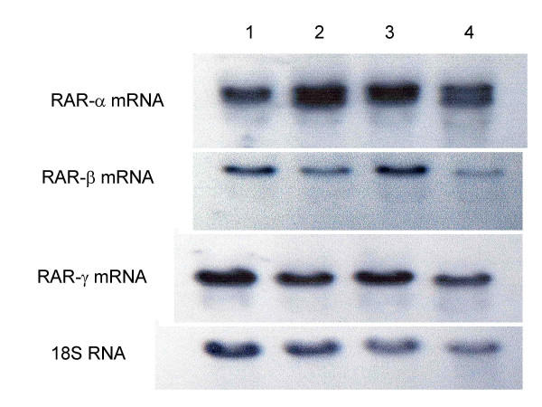
Results: We observed that prostatic cell lines stably expressing S3c required STAT3 expression for survival, because they became sensitive to antisense oligonucleotide for STAT3. However, S3c-transfected cells were not sensitive to the effects of JAK inhibitors, meaning that STAT3 was constitutively-activated in these transfected cell lines. NRP-152 prostatic epithelial cells lost the requirement for exogenous growth factors. Furthermore, we observed that NRP-152 expressing S3c had enhanced mRNA levels of retinoic acid receptor (RAR)-alpha, reduced mRNA levels of RAR-beta and -gamma, while BPH-1 cells transfected with S3c became insensitive to the effects of androgen, and also to the effects of a testosterone antagonist. Both S3c-transfected cell lines grew in soft agar after stable transfection with S3c, however neither S3c-transfected cell line was tumorigenic in severe-combined immunodeficient mice.

Conclusions: We conclude, based on our findings, that persistently-activated STAT3 is an important molecular marker of prostate cancer, which develops in formerly benign prostate cells and changes their phenotype to one more closely resembling transformed prostate cells. That the S3c-transfected cell lines require the continued expression of S3c demonstrates that a significant phenotypic change occurred in the cells. These conclusions are based on our data with respect to loss of growth factor requirement, loss of androgen response, gain of growth in soft agar, and changes in RAR subunit expression, all of which are consistent with a malignant phenotype in prostate cancer. However, an additional genetic change may be required for S3c-transfected prostate cells to become tumorigenic.


Figures

Figure 1
Figure 1
An example of cytokine-mediated activation of STAT3. In this example, IL-6-induced binding to its receptor leads to homodimerization of the receptor, which in turn leads to autophosphorylation of the cytosolic domain of gp130; this in turn causes the phosphorylation of one of 3 kinases, JAK1, JAK2, or Tyk 2. The activated up-stream kinase phosphorylates STAT3, which allows for dimerization of STAT3; only the dimer can translocate and dock to DNA at target genes, thereby directing transcription.
Figure 2
Figure 2
FLAG and EGFP expression in representative NRP-152 and BPH-1 clones transfected with either pBABE-S3c or pIRES-S3c. NRP-152 and BPH-1 cells were transfected with pBABE-S3c or pIRES-S3c, which bear the FLAG epitope on the S3c gene. Clones were derived by limit dilution, as described in Materials & Methods. Panels A–D: In all histograms, the marker M1 sets the region of positively fluorescent cells for determining the percent positive cells. Panels A & B: Fixed cells were permeabilized and stained with anti-FLAG M1 Ab (Sigma), as described in Materials & Methods. Controls for staining were included, as described. Panel A: Transfected NRP-152 cells. Thin line = 152-S3c; thick line = NRP-152. Panel B: Transfected BPH-1 cells. Thin line = BPH-1; thick line = BPH-S3c. Panels C & D: NRP-152 and BPH-1 cells transfected with pIRES-S3c were analyzed for EGFP fluorescence, following selection. Panel C: Transfected NRP-152 cells. Thin line = NRP-152; thin line = 152-S3c. Panel D: Transfected BPH-1 cells. Thick line = BPH-1; thin line = BPH-S3c. Panel E: Immunoprecipitation followed by Western blot showing EGFP expression in transfected NRP-152 and BPH cells. Note the lack of EGFP bands for parental lines NRP-152 and BPH-1, whereas EGFP was detected using EGFP-specific Ab (Pharmingen) in lanes for 152-S3c and BPH-S3c. Methods: NRP-152, 152-S3c, BPH-1, and BPH-S3c cells were lysed in buffer. Equal amounts of protein in cell lysates were pre-cleared with Protein A/G beads, then precipitated with anti-FLAG AB plus Protein A/G beads with rotation in the cold. The pelleted beads plus proteins were separated on 12% SDS gels, transferred to PVDF membranes, then blotted with Ab to EGFP. Enhanced chemifluorescence was used to reveal the 27 kD bands corresponding to EGFP.
Figure 3
Figure 3
Growth of NRP-152, NRP-154, 152pBABE, and 152-S3c clones on 154 medium compared to growth on 152 medium. 103 cells were seeded in microtiter wells, in the indicated medium. After incubation for 48 hr, MTT (15 μl at 25 μg/ml) were added to each well, and incubation was continued for 4 hr more. The formazan was dissolved in 0.1% SDS, and the absorbance was quantified on a DynaTech plate reader at 570 nm. Unpaired Student t-tests (InStat3 software) were performed to assess the statistical significance of the growth of S3c-transfected cells relative to pBABE transfected and untransfected NRP-152 cells. Panel A: Comparison of growth as measured by MTT absorbance at 48 hours; Panel B: Comparison of growth rates over 72 hours.
Figure 4
Figure 4
Functional activity of STAT3 in S3c-transfected cells. Panel A: To show the functional activity of STAT3 expressed by the S3c gene, BPH-1 cells stably transfected with pIRES-S3c were treated with either 125 nM sense or antisense STAT3 oligonucleotide. Percent viability over time was determined by staining with propidium iodide, then quantifying fluorescence on a FACScan flow cytometer. Panel B: Treatment with 125 nM antisense STAT3 oligo reduced the amount of intracellular STAT3 protein in the clone of BPH-S3c cells shown in 3A. Twenty-four hours after transfection, BPH-S3c cells were harvested, fixed, and permeabilized, then stained with antibody to STAT3, as described in Materials and Methods. Quantification was performed on a FACScan flow cytometer. The black line indicates the amount of intracellular STAT3 in BPH-S3c cells treated with sense STAT3, while the grey line shows the amount of STAT3 in BPH-S3c cells given antisense STAT3. STAT3 expression was reduced by 66% in this experiment.
Figure 5
Figure 5
S3c expression inhibited RAR-β and -γ expression and increased expression of RAR-α in NRP-152 cells. Panel A: Effect of S3c on RAR-β mRNA levels. Panel B: Effect of S3c on RAR-γ mRNA levels. Panel C: Effect of S3c on RAR-α mRNA levels. NRP-152, NRP-154, NRP-pBABE, and 152-S3c cells were grown to confluence, and RNA was harvested as described in Materials & Methods. Electrophoretic separation of RNA was followed by transfer to nitrocellulose, then hybridization with 32P-labeled probe, followed by autoradiography. Lane 1 = NRP-152 (rat benign prostatic hyperplasia line); lane 2 = NRP-154 (rat prostatic carcinoma line); lane 3 = 152-pBABE; lane 4 = 152-S3c. The comparison to 18S RNA is shown for each.

Similar articles

Cited by

References

    1. Ihle JN. STATs and MAPKs: obligate or opportunistic partners in signaling. BioEssays. 1996;18:95–98. - PubMed
    1. Aaronson DS, Horvath CM. A Road Map for Those Who Don't Know JAK-STAT. Science. 2002;296:1653–1655. doi: 10.1126/science.1071545. - DOI - PubMed
    1. Schroder M, Kroeger KM, Volk HD, Eidne KA, G. G. Preassociation of nonactivated STAT3 molecules demonstrated in living cells using bioluminescence resonance energy transfer: a new model of STAT activation? J Leukoc Biol. 2004;75:792–797. doi: 10.1189/jlb.1003496. - DOI - PubMed
    1. Kretzschmar AK, Dinger MC, Henze C, Brocke-Heidrich K, Horn F. Analysis of Stat3 (signal transducer and activator of transcription 3) dimerization by fluorescence resonance energy transfer in living cells. Biochem J. 2004;377:289–297. doi: 10.1042/BJ20030708. - DOI - PMC - PubMed
    1. Sehgal PB. Plasma membrane rafts and chaperones in cytokine/STAT signaling. Acta Biochim Pol. 2003;50:583–594. - PubMed

Publication types

MeSH terms