. 2005 Jan 12:6:4.
doi: 10.1186/1471-2164-6-4.

Structure and evolution of the mouse pregnancy-specific glycoprotein (Psg) gene locus

Affiliations

Structure and evolution of the mouse pregnancy-specific glycoprotein (Psg) gene locus

Andrew S McLellan et al. BMC Genomics. .

Abstract

Background: The pregnancy-specific glycoprotein (Psg) genes encode proteins of unknown function, and are members of the carcinoembryonic antigen (Cea) gene family, which is a member of the immunoglobulin gene (Ig) superfamily. In rodents and primates, but not in artiodactyls (even-toed ungulates / hoofed mammals), there have been independent expansions of the Psg gene family, with all members expressed exclusively in placental trophoblast cells. For the mouse Psg genes, we sought to determine the genomic organisation of the locus, the expression profiles of the various family members, and the evolution of exon structure, to attempt to reconstruct the evolutionary history of this locus, and to determine whether expansion of the gene family has been driven by selection for increased gene dosage, or diversification of function.

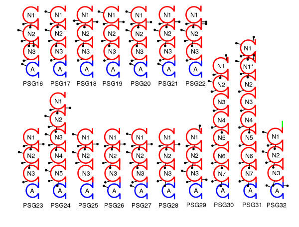
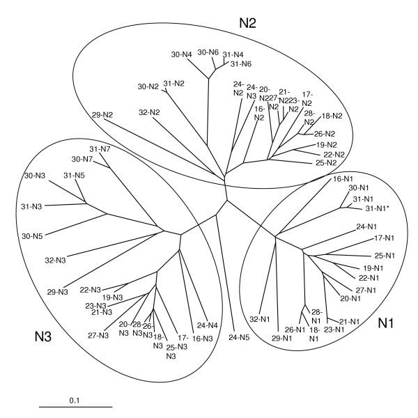
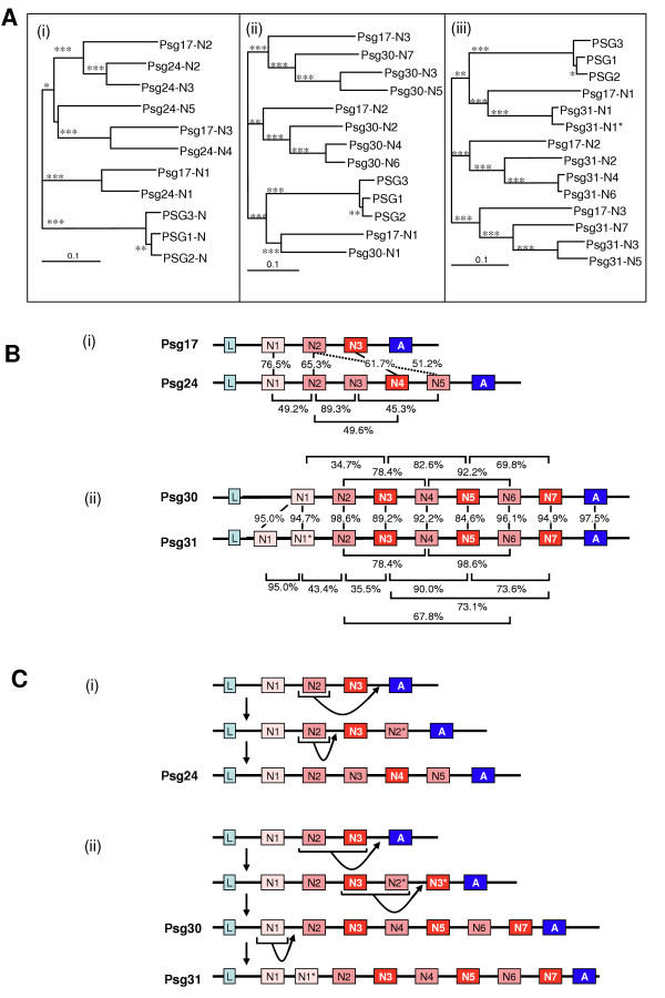
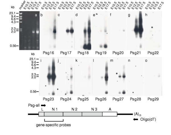
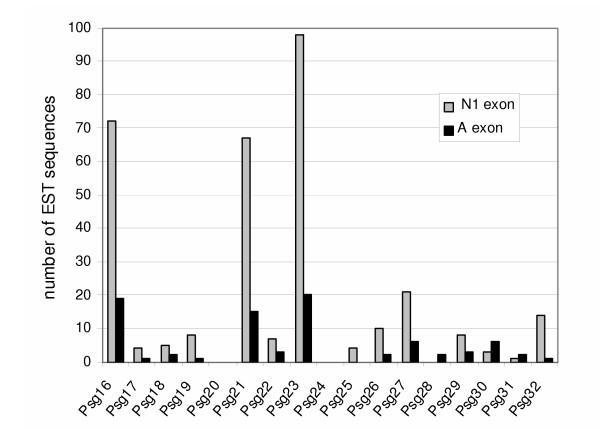
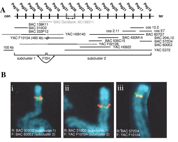
Results: We collated the mouse Psg gene sequences currently in the public genome and expressed-sequence tag (EST) databases and used systematic BLAST searches to generate complete sequences for all known mouse Psg genes. We identified a novel family member, Psg31, which is similar to Psg30 but, uniquely amongst mouse Psg genes, has a duplicated N1 domain. We also identified a novel splice variant of Psg16 (bCEA). We show that Psg24 and Psg30 / Psg31 have independently undergone expansion of N-domain number. By mapping BAC, YAC and cosmid clones we described two clusters of Psg genes, which we linked and oriented using fluorescent in situ hybridisation (FISH). Comparison of our Psg locus map with the public mouse genome database indicates good agreement in overall structure and further elucidates gene order. Expression levels of Psg genes in placentas of different developmental stages revealed dramatic differences in the developmental expression profile of individual family members.

Conclusion: We have combined existing information, and provide new information concerning the evolution of mouse Psg exon organization, the mouse Psg genomic locus structure, and the expression patterns of individual Psg genes. This information will facilitate functional studies of this complex gene family.


Figures

Figure 1
Figure 1
Domain organization of mouse PSGs. Mouse PSGs are composed of 3 – 8 IgV-like N domains and one IgC-like A domain. The relative position of potential N-glycosylation sites (consensus amino acid sequence: asparagine-X-threonine / serine; X any amino acid except proline) were identified using the NetNGlyc 1.0 Server online software and indicated by lollipops. Although PSG32 is probably not routed through the endoplasmic reticulum, the putative N-glycosylation sites are shown for comparison. Of the two PSG16 splice variants, only the variant expressed in the placenta is shown.
Figure 2
Figure 2
Evolutionary relationships between mouse PSG IgV-like domains. An unrooted evolutionary tree based on ClustalX amino acid sequence alignments showing the relationships between all mouse PSG N-domains. The three main groups N1, N2 and N3 have been ringed for clarity. The scale bar represents 0.1 amino acid substitutions per site.
Figure 3
Figure 3
Domain expansion of Psg24, Psg30 and Psg31. A. NJ-trees based on ClustalX amino acid sequence alignments showing: (i) the evolution of PSG24 IgV-like domains compared to those of PSG17; (ii) the evolution of PSG30 IgV-like domains compared to those of PSG17; (iii) the evolution of PSG31 IgV-like domains compared to those of PSG17. The trees were rooted using an outgroup consisting of the N-domain amino acid sequences of human PSG1, PSG2 and PSG3. Alignments were bootstrapped 1000 times yielding node values which are represented as follows < 50%: no mark; 50–74%: marked *; 75–94%: marked **; ≥ 95%: marked ***. The scale bar represents 0.1 amino acid substitutions per site. B. The arrangement of domains represented by boxes shaded: cyan for leader (L) peptides; light pink for the N1-domains; dark pink for N2 and N2-like domains; red for N3 and N3-like domains; blue for A-domains. (i) Comparison of Psg17 and Psg24 exon arrangement including identities of amino acid sequence alignments. (ii) Comparison of Psg30 and Psg31 exon arrangements including identities of amino acid sequence alignments. C. Predicted model of IgV-like domain expansion by exon duplications in (i) Psg24 and (ii) Psg30 and Psg31.
Figure 4
Figure 4
Expression of Psg mRNAs during placental development. Total RNA (1 μg) from day 10.5, 12.5, 15.5 and 17.5 BALB/c placentae was reverse transcribed using an oligo (dT) oligonucleotide (reverse PCR primer). After addition of the degenerate Psg-all oligonucleotide (forward PCR primer), which anneals to the cDNA of all known members of the mouse Psg family, Psg cDNAs were amplified by PCR (see schematic diagram depicting generalised mouse Psg cDNA amplification). Aliquots were size-separated by agarose gel electrophoresis. a, PCR products were visualised by ethidium bromide staining. b-o, the amplification products were blotted onto nylon membranes and individual blots were hybridised with single gene-specific 32P-labelled oligonucleotides from the N1 domain regions (Table 2). The location of the primers used for amplification of the Psg cDNAs and the region from which the sequences of the gene-specific oligonucleotides were derived are shown together with a schematic representation of mouse Psg mRNA. The 5'- and 3'-untranslated regions are shown as bold lines. L, leader; N1-N3, IgV-like domains; A, IgC-like domain.
Figure 5
Figure 5
Virtual Northern analysis of the mouse Psg genes. The nucleotide sequences of the Psg exons encoding the N1 or the A domains were used in NCBI-BLAST searches of the GenBank mouse EST database (March 16, 2004) for the presence of Psg transcripts (virtual Northern analysis). A hit was registered when a 100% match for a sequence > 150 bp was observed. Obvious mismatches such as unidentified nucleotides (N) or single nucleotide insertions or deletions (especially at the end of a sequence run) were ignored.
Figure 6
Figure 6
Physical map of mouse Psg gene locus. A. The order of the Psg genes was inferred from the presence of the various genes on overlapping cosmid, BAC and YAC clones. The position of Psgs represented by filled boxes is unequivocal, whereas the position of those represented by open boxes is ambiguous. Arrows between pairs of genes indicate that their order remains unresolved. The distances between individual genes are not shown to scale. Chimeric YACs mapping to separate chromosomes are indicated by stippled and solid lines. The solid lines correspond to chromosome 7 regions containing the Psg genes indicated above. The locations of the non-chromosome 7 regions are not known. Only the sizes of non-chimeric YACs have been determined and are shown (size bar corresponds to 100 kb). The centromere (cen) / telomere (ter) order and the relative orientation of the two Psg gene subclusters were resolved by FISH mapping. B. Two-colour FISH prophase mapping of relative orientation of the two Psg gene subclusters using mouse m5S cells and C57BL/6CrSlc mouse lymphocytes. (i) FISH pattern representative of 38 experiments where BAC 310D2 in subcluster 1, labelled with rhodamine (R), is centromeric to BAC 600E2 from subcluster 2, labelled with fluorosceine (F). (ii) FISH pattern representative of 38 experiments where BAC 310D2 in subcluster 1, labelled with rhodamine, is centromeric to YAC F10104 from subcluster 2, labelled with fluorosceine. (iii) Orientation of subcluster 2 determined by relative positions of BAC 572D4, labelled with rhodamine, which is telomeric to YAC F10104, labelled with fluorosceine.

References

    1. Soares MJ. The prolactin and growth hormone families: pregnancy-specific hormones/cytokines at the maternal-fetal interface. Reproductive Biology and Endocrinology. 2004;2:51. doi: 10.1186/1477-7827-2-51. - DOI - PMC - PubMed
    1. Lin TM, Halbert SP, Spellacy WN. Measurement of pregnancy-associated plasma proteins during human gestation. Journal of Clinical Investigation. 1974;54:576–582. - PMC - PubMed
    1. Lei KJ, Sartwell AD, Pan CJ, Chou JY. Cloning and expression of genes encoding human pregnancy-specific glycoproteins. Journal of Biological Chemistry. 1992;267:16371–16378. - PubMed
    1. Rebstock S, Lucas K, Weiss M, Thompson J, Zimmermann W. Spatiotemporal expression of pregnancy-specific glycoprotein gene rnCGM1 in rat placenta. Developmental Dynamics. 1993;198:171–181. - PubMed
    1. Kromer B, Finkenzeller D, Wessels J, Dveksler G, Thompson J, Zimmermann W. Coordinate expression of splice variants of the murine pregnancy-specific glycoprotein (PSG) gene family during placental development. European Journal of Biochemistry. 1996;242:280–287. doi: 10.1111/j.1432-1033.1996.0280r.x. - DOI - PubMed

Publication types

MeSH terms