

Histological precursors of oesophageal squamous cell carcinoma: results from a 13 year prospective follow up study in a high risk population
- PMID: 15647178
- PMCID: PMC1774842
- DOI: 10.1136/gut.2004.046631
Histological precursors of oesophageal squamous cell carcinoma: results from a 13 year prospective follow up study in a high risk population
Abstract
Background: Oesophageal squamous cell carcinoma (OSCC) has a very poor prognosis, which is largely due to late diagnosis. Successful early detection strategies will require identification of clinically relevant precursor lesions that can be targets for screening and treatment.
Aims: To identify the clinically relevant histological precursors of OSCC.
Subjects: A cohort of 682 endoscoped patients from a high risk rural population in Linxian, China.
Methods: Subjects were endoscoped and biopsied at baseline and followed for 13.5 years. We estimated the relative risk of developing OSCC for each of the initial histological diagnoses using Cox proportional hazards regression models.
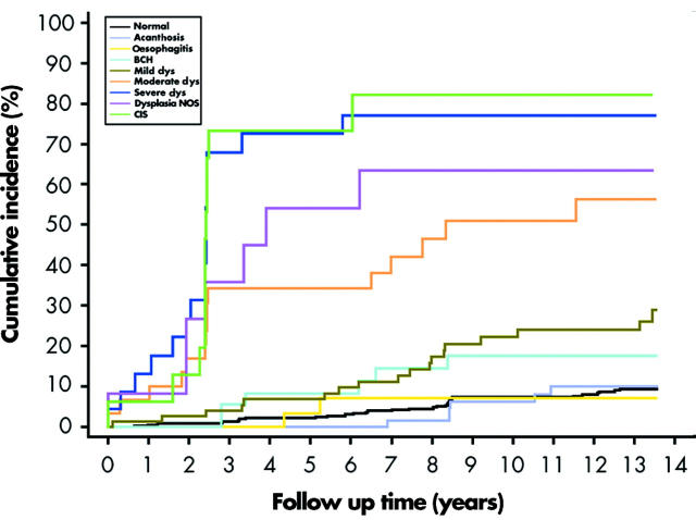
Results: A total of 114 (16.7%) patients developed OSCC during the follow up period. After adjusting for potential confounding factors, relative risks (95% confidence intervals) for incidence of this tumour, by initial histological diagnosis, were: normal 1.0 (reference), oesophagitis 0.8 (0.2-3.2), basal cell hyperplasia 1.9 (0.8-4.5), mild dysplasia 2.9 (1.6-5.2), moderate dysplasia 9.8 (5.3-18.3), severe dysplasia 28.3 (15.3-52.3), and carcinoma in situ 34.4 (16.6-71.4).
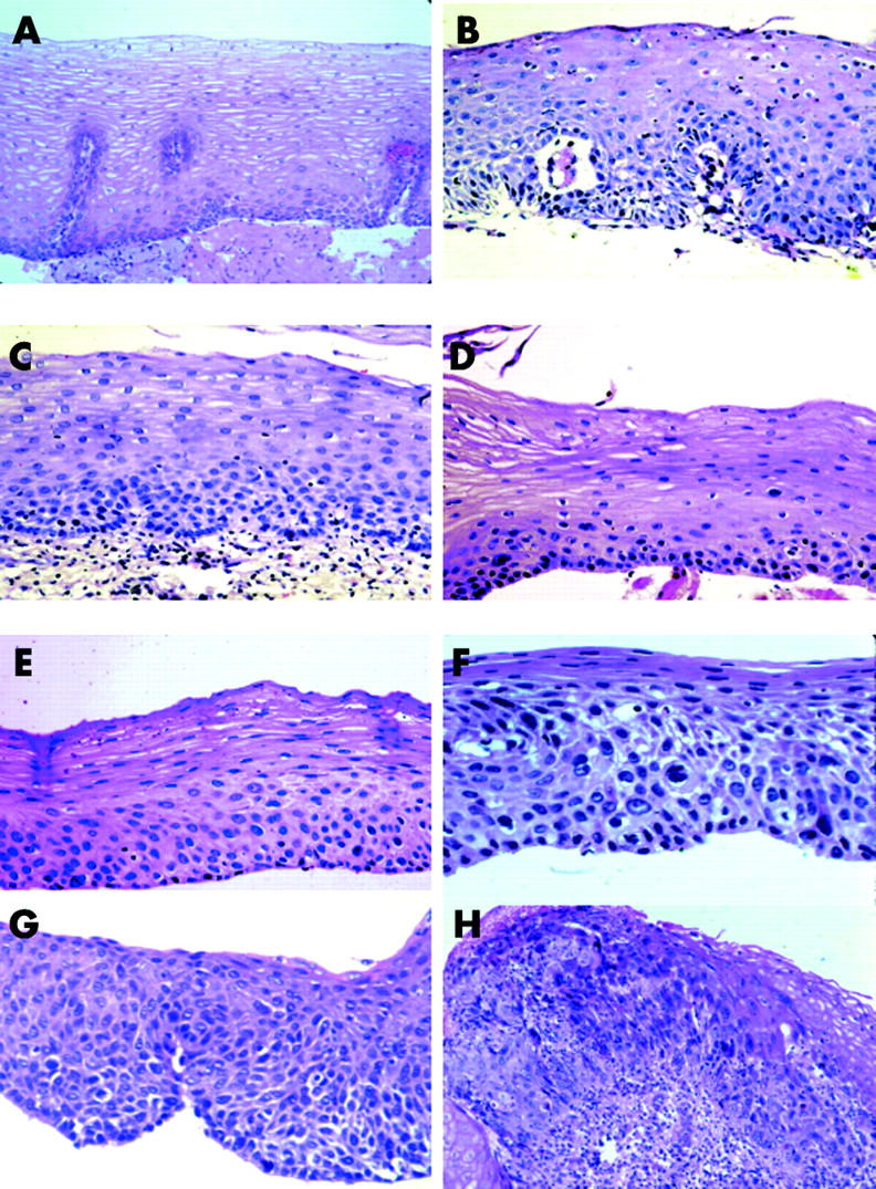
Conclusions: In this study, squamous dysplasia and carcinoma in situ were the only histological lesions associated with a significantly increased risk of developing OSCC within 13.5 years after endoscopy. There was no evidence that oesophagitis predisposed to this tumour. Increasing grades of dysplasia were strongly associated with increasing risk, indicating that the histological grading was clinically meaningful. The follow up experience of severe dysplasia and carcinoma in situ was equivalent, suggesting that this distinction is not clinically relevant. Documenting these precursor lesions of OSCC should assist in the development of effective prevention, early detection, and treatment strategies for this disease.
Figures
References
-
- Parkin DM, Bray FI, Devesa SS. Cancer burden in the year 2000. The global picture. Eur J Cancer 2001;37 (suppl 8) :S4–66. - PubMed
-
- Ries LAG, Eisner MP, Kosary CL, et al. eds. SEER cancer statistics review, 1975–2000. Bethesda, MD: National Cancer Institute, 2003.
-
- Munoz N , Day NE. Esophageal cancer. In: Schottenfeld D, Fraumeni JF Jr, eds. Cancer epidemiology and prevention. New York: Oxford University Press, 1996:681–706.
-
- Blot WJ, Li J-Y, Taylor PR, et al. Nutrition intervention trials in Linxian, China: supplementation with specific vitamin/mineral combinations, cancer incidence, and disease-specific mortality in the general population. J Natl Cancer Inst 1993;85:1483–92. - PubMed
-
- Li B , Taylor PR, Li J-Y, et al. Linxian nutrition intervention trials. Design, methods, participant characteristics, and compliance. Ann Epidemiol 1993;3:577–85. - PubMed
Publication types
MeSH terms
Grants and funding
LinkOut - more resources
Full Text Sources
Medical