. 2005 Feb;54(2):257-63.
doi: 10.1136/gut.2003.037085.

Magnetic resonance colonography for the detection of inflammatory diseases of the large bowel: quantifying the inflammatory activity

Affiliations

Magnetic resonance colonography for the detection of inflammatory diseases of the large bowel: quantifying the inflammatory activity

W M Ajaj et al. Gut. 2005 Feb.

Abstract

Introduction: The purpose of this study was to assess the diagnostic accuracy of magnetic resonance colonography (MRC) for its ability to detect and quantify inflammatory bowel disease (IBD) affecting the colon. Endoscopically obtained histopathology specimens were used as the standard of reference.

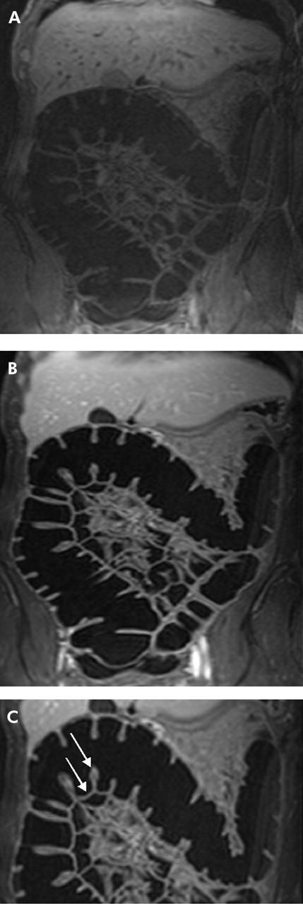
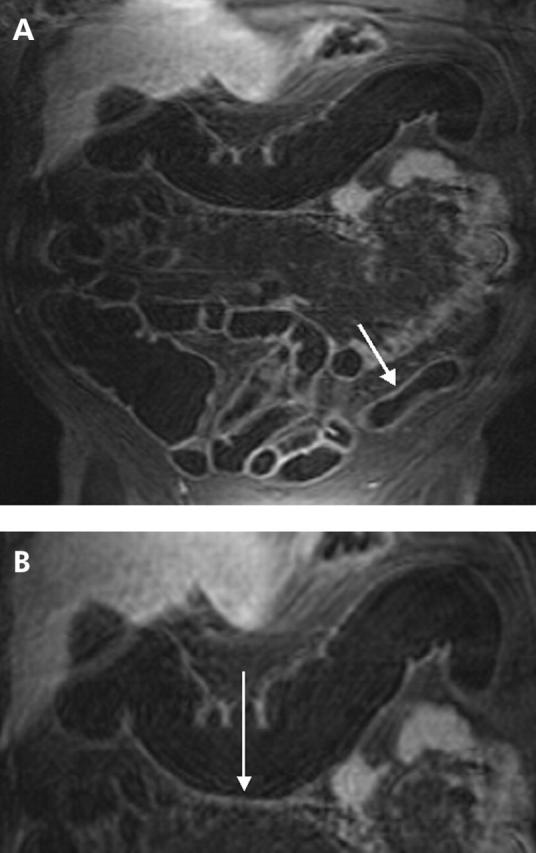
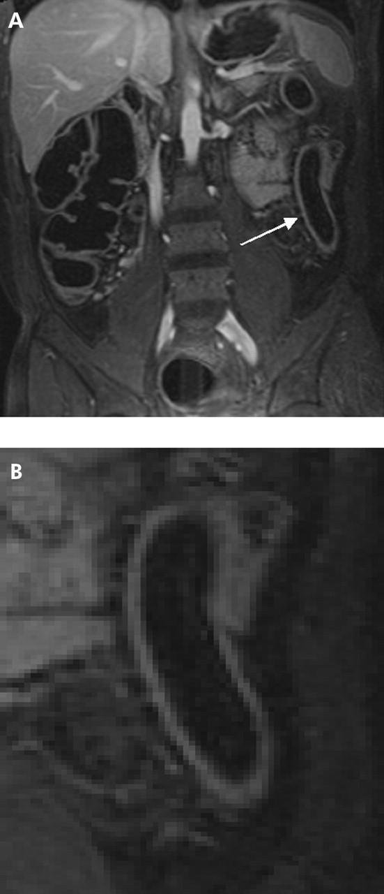
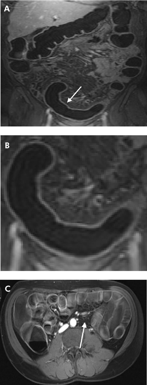
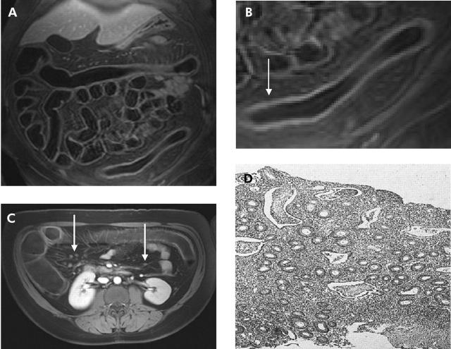
Materials and methods: Fifteen normal subjects and 23 patients with suspected IBD of the large bowel underwent MRC. Three dimensional T1 weighted data sets were collected following rectal administration of water prior to and 75 seconds after intravenous administration of paramagnetic contrast (gadolinium-BOPTA). The presence of inflammatory changes in patients was documented based on bowel wall contrast enhancement, bowel wall thickness, presence of perifocal lymph nodes, and loss of haustral folds. All four criteria were quantified relative to data obtained from normal subjects and summarised in a single score. This MRC based score was compared with histopathological data based on conventional endoscopic findings.

Results: MRC correctly identified 68 of 73 segments found to reveal IBD changes by histopathology. All severely inflamed segments were correctly identified as such and there were no false positive findings. Based on the proposed composite score, MRC detected and characterised clinically relevant IBD of the large bowel with sensitivity and specificity values of 87% and 100%, respectively, for all investigated colonic segments.

Conclusion: MRC may be considered a promising alternative to endoscopic biopsy in monitoring IBD activity or assessing therapeutic effectiveness.


Figures

Figure 1
Figure 1
(A) A 38 year old volunteer undergoing magnetic resonance colonography in conjunction with rectal application of water. The coronal source image of T1 weighted three dimensional GRE (TR/TE 3.1/1.1) scan acquired prior to intravenous application of contrast medium shows moderate contrast between bowel wall and bowel lumen. (B) Coronal source images of the same volunteer acquired 75 seconds after intravenous administration of gadolinium. The colonic wall enhances brightly and can be easily delineated against the background of a dark water filled colonic lumen. (C) Transverse colon of the same volunteer. The colonic wall shows a normal thickness (2 mm) (arrow), contrast uptake (contrast to noise ratio 42), and number of haustral folds (14) (arrow).
Figure 2
Figure 2
(A) T1 weighted three dimensional GRE image (TR/TE 3.1/1.1) of a 55 year old male patient with known Crohn’s disease. Magnetic resonance colonography (MRC) visualised a slight inflammation in the descending colon based on an increase in contrast uptake and wall thickness and decrease in the number of haustral folds (arrow). This finding was confirmed by histopathology. (B) Detailed display of the transverse colon of the same patient. By means of MRC, the transverse colon was graded as normal (arrow). However, histopathology showed slight inflammation in the transverse colon.
Figure 3
Figure 3
(A) T1 weighted three dimensional GRE image (TR/TE 3.1/1.1) of a 47 year old male patient with known ulcerative colitis. Magnetic resonance colonography diagnosed an inflammation in the descending colon (arrow). (B) Detailed display of the descending colon of the same patient which displays loss of haustral markings and slight bowel wall thickening. Due to the absence of lymph nodes as well as normal contrast uptake in the colonic wall, inflammation was rated as slight. This was confirmed by subsequent endoscopy.
Figure 4
Figure 4
(A) T1 weighted three dimensional GRE image (TR/TE 3.1/1.1) of 31 year old male patient with known Crohn’s disease. An inflammatory process was detected in the rectum and sigmoid colon (arrow). (B) Detailed display of (A). Loss of haustral markings and increased contrast uptake of the colonic wall as well as bowel wall thickening were determined leading to a diagnosis of inflammation. (C) On the axial reformatted image, several mesenteric lymph nodes were found (arrow). Subsequent endoscopy and biopsy confirmed the presence of an acute moderate inflammation of the sigmoid colon.
Figure 5
Figure 5
(A) T1 weighted three dimensional GRE image (TR/TE 3.1/1.1) of a 39 year old female patient with known ulcerative colitis. (B) Detailed display of the descending colon of (A). Complete loss of haustral markings, increased contrast uptake of the colonic wall, as well as bowel wall thickening were determined (arrow). (C) Axial reformatted T1 weighted image of the same patient. Multiple mesenteric lymph nodes were found (arrows). (D) Histopathology of the descending colon confirmed the diagnosis of an acute severe ulcerative colitis showing loss of haustral fold, multiple crypt abscesses, and infiltration with granulocytes.

Comment in

Similar articles

Cited by

References

    1. Kiesslich R , Jung M, Galle PR, et al. Ulcerative colitis—how can endoscopic observation be improved? Dtsch Med Wochenschr 2003;128:139–41. - PubMed
    1. Fiocca R , Ceppa P. The diagnostic reliability of endoscopic biopsies in diagnosis colitis. J Clin Pathol 2003;56:321–2. - PMC - PubMed
    1. Nahon S , Bouhnik Y, Lavergne-Slove A, et al. Colonoscopy accurately predicts the anatomical severity of colonic Crohn’s disease attacks: correlation with findings from colectomy specimens. Am J Gastroenterol 2002;12:3102–7. - PubMed
    1. Eckardt VF, Gaedertz C, Eidner C. Colonic perforation with endoscopic biopsy. Gastrointest Endosc 1997;6:560–2. - PubMed
    1. Cappell MS, Friedel D. The role of sigmoidoscopy and colonoscopy in the diagnosis and management of lower gastrointestinal disorders: endoscopic findings, therapy, and complications. Med Clin North Am 2002;6:1253–88. - PubMed

Publication types