. 2005 Apr 8;280(14):13779-83.
doi: 10.1074/jbc.M413781200. Epub 2005 Jan 12.

Crystal structure of the virulence gene activator AphA from Vibrio cholerae reveals it is a novel member of the winged helix transcription factor superfamily

Affiliations

Crystal structure of the virulence gene activator AphA from Vibrio cholerae reveals it is a novel member of the winged helix transcription factor superfamily

Rukman S De Silva et al. J Biol Chem. .

Abstract

AphA is a member of a new and largely uncharacterized family of transcriptional activators that is required for initiating virulence gene expression in Vibrio cholerae, the causative agent of the frequently fatal epidemic diarrheal disease cholera. AphA activates transcription by an unusual mechanism that appears to involve a direct interaction with the LysR-type regulator AphB at the tcpPH promoter. As a first step toward understanding the molecular basis for tcpPH activation by AphA and AphB, we have determined the crystal structure of AphA to 2.2 angstrom resolution. AphA is a dimer with an N-terminal winged helix DNA binding domain that is architecturally similar to that of the MarR family of transcriptional regulators. Unlike this family, however, AphA has a unique C-terminal antiparallel coiled coil domain that serves as its primary dimerization interface. AphA monomers are highly unstable by themselves and form a linked topology, requiring the protein to partially unfold to form the dimer. The structure of AphA also provides insights into how it cooperates with AphB to activate transcription, most likely by forming a heterotetrameric complex at the tcpPH promoter.


Figures
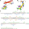
FIG. 1
FIG. 1. Structure of AphA and comparison with MarR
A, side view of the AphA monomer showing secondary structural elements. Unresolved residues in the wing motif are shown as a dashed line. B, side view of MarR (Protein Data Bank number 1JGS) in the same orientation. The analogous helices are similarly color coded. The N-terminal helix α1 of MarR that is absent in AphA is shown in dark blue. The MarR helix α5 (yellow) is much longer than AphA α4 and forms the main dimerization interface. The shorter MarR helix α6 (orange) has only a minor role in dimerization. C, sequence alignment of AphA and MarR. Secondary structural elements, as determined from the crystal structures, are shown above and below the sequences (the first six residues of MarR were disordered). H, α helices; S, β-strands; ., residues not visualized in the crystal structure. Identical residues between the two are shown in red. Bold characters in the AphA sequence show positions of mutations described in the text that influence its DNA binding and dimerization activities. Homologous α-carbons within the DNA binding domain that superpose according to the program DALI (18) include: AphA (amino acids 1–14) with MarR (Amino acids 33–46), AphA (amino acids 15–26) with MarR (amino acids 48–59), AphA (amino acids 35–83) with MarR (amino acids 60–108), AphA (amino acids 84–87) with MarR (amino acids 111–14), and AphA (amino acids 88–91) with MarR (amino acids 117–20).
FIG. 2
FIG. 2. Structure of the AphA and MarR dimers
A, the AphA dimer viewed along the crystallographic two-fold axis. In this top view, the DNA binding face is underneath the dimer. For one molecule, the N-terminal DNA binding domain is green, and the C-terminal dimerization domain is red. For the second molecule, the DNA and dimerization domains are blue and orange, respectively. Unresolved residues in the wing motif are shown as a dashed line. B, electrostatic surface potential of the AphA dimer, oriented as in A. The complementary positive (blue) and negative (red) charge clusters that stabilize the dimer interface can be seen at both ends of the α7 helices. C, the AphA dimer viewed from the side, colored as in A. The DNA binding surface is at the bottom in this orientation. D, side view of the MarR dimer. The DNA binding and dimerization domains are colored the same as for AphA.
FIG. 3
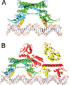
FIG. 3. Models of protein·DNA complexes
A, a model of the AphA dimer (one chain in blue and the other in green) bound to its DNA site with the complete AphA wing modeled in, based on the wing structure observed in MarR. The primary DNA binding interface is predicted to involve helix α3 from each monomer (orange), which fit into adjacent major grooves on the same face of the DNA. As the helices are not optimally spaced, some conformational change in the protein and/or DNA likely takes place. B, model of the AphA-AphB heterotetramer bound to DNA. The AphB dimer structure was modeled by threading the AphB sequence into the crystal structure of CbnR and then placing it next to the AphA-DNA model shown in A. In this orientation, the C-terminal domain of one molecule of AphB (red) would be able to interact with the AphA dimer (green and blue). Interaction between AphA and AphB could also form between the tips of the wings. This model predicts a linear mode of binding that would not significantly distort the DNA in which four adjacent major grooves are bound by the AphA dimer and the AphB dimer.

Similar articles

Cited by

References

    1. Taylor RK, Miller VL, Furlong DB, Mekalanos JJ. Proc. Natl. Acad. Sci. U. S. A. 1987;84:2833–2837. - PMC - PubMed
    1. Kaper JB, Morris JG, Jr, Levine MM. Clin. Microbiol. Rev. 1995;8:48–86. - PMC - PubMed
    1. Karaolis DK, Johnson JA, Bailey CC, Boedeker EC, Kaper JB, Reeves PR. Proc. Natl. Acad. Sci. U. S. A. 1998;95:3134–3139. - PMC - PubMed
    1. Waldor MK, Mekalanos JJ. Science. 1996;272:1910–1914. - PubMed
    1. Skorupski K, Taylor RK. Mol. Microbiol. 1999;31:763–771. - PubMed

Publication types

Associated data

LinkOut - more resources