. 2005 Mar;16(3):1406-16.
doi: 10.1091/mbc.e04-10-0920. Epub 2005 Jan 12.

Organelle-specific control of intracellular transport: distinctly targeted isoforms of the regulator Klar

Affiliations

Organelle-specific control of intracellular transport: distinctly targeted isoforms of the regulator Klar

Yi Guo et al. Mol Biol Cell. 2005 Mar.

Abstract

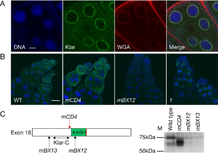
Microtubule-based transport in cells is powered by a small set of distinct motors, yet timing and destination of transport can be controlled in a cargo-specific manner. The mechanistic basis for this specificity is not understood. To address this question, we analyzed the Drosophila Klarsicht (Klar) protein that regulates distinct microtubule-based transport processes. We find that localization of Klar to its cargoes is crucial for Klar function. Using mutations, we identify functionally important regions of Klar that confer distinct cargo specificity. In ovaries, Klar is present on the nuclear envelope, a localization that requires the C-terminal KASH domain. In early embryos, Klar is attached to lipid droplets, a localization mediated by a novel C-terminal domain encoded by an alternatively spliced exon. In cultured cells, these two domains are sufficient for targeting to the correct intracellular location. Our analysis disentangles Klar's modular organization: we propose that a core region integral to motor regulation is attached to variable domains so that the cell can target regulators with overlapping, yet distinct functions to specific cargoes. Such isoform variation may be a general strategy for adapting a common regulatory mechanism to specifically control motion and positioning of multiple organelles.


Figures

Figure 1.
Figure 1.
Molecular lesions in various klar alleles and their effect on Klar protein expression in early embryos. (A) The klar gene spans ∼100 kb in the genome. This map is based on the klar gene annotation available on FlyBase (The FlyBase Consortium, 2003). Predicted exons are labeled from 0 to 18. Translation is predicted to start in exon 2 (cyan line) and stop in exon 18 (red line). The green box in exon 18 indicates the KASH domain. The epitopes of antibodies Klar-N, Klar-M, and Klar-C were mapped to exon 4, 9, and 18, respectively, as indicated by double-headed arrows and the corresponding amino acid number. (B and C) Proteins from phase II embryos of various genotypes were separated by SDS-PAGE and analyzed by Western blotting by using antibody Klar-M (B) or Klar-N (C). Both antibodies detect a major form of Klar above 250 kDa, plus additional bands. Comparison to alleles with N-terminal lesions (e.g., klarB) shows that all these bands are Klar specific. These bands might be breakdown products of a longer form or specific Klar isoforms resulting from alternative splicing/protein processing. Class II alleles express Klar proteins similar in size to the wild type. Class I alleles express truncated protein forms. (D) Molecular lesions in various alleles. The klar gene structure is the same as in A. Arrows point to the approximate position of the molecular lesions of klar alleles (see Table 1 for details). Alleles with nonsense mutations are red, and alleles with chromosomal breaks are blue. Lesions of class I alleles are located 5′ to exon 15, whereas class II alleles disrupt exon 18.
Figure 3.
Figure 3.
Motion of lipid droplets and of photoreceptor nuclei require partially distinct and partially overlapping regions of Klar. Class I alleles (represented by klar1) disrupt droplet transport, whereas class II alleles (represented by klarmBX12) do not. Both classes disrupt nuclear migration in photoreceptor cells. (A–I) Wild-type, class I, and class II embryos were compared by transmitted light to reveal changes in transparency (A–C, phase II; D–F, phase III) or by staining with Nile Red to detect lipid droplets (G–I, phase III, green). In phase III embryos, lipid droplets remain basally for class I alleles but spread apically in the other genotypes. (J–L) Wild-type, klar1, and klarmBX12 eye imaginal disks were fixed and stained for the neural antigen Elav (green) to reveal photoreceptor nuclei. Distribution of nuclei at the apical side of the disk was documented by confocal microscopy. In the wild type, the nuclei are arranged in a regular pattern apically. For both klar alleles, most nuclei are missing from this apical region, indicating basal mislocalization. Bars, 100 μm (A–I) and 8 μm (J–L).
Figure 4.
Figure 4.
Alternate exons of klar. (A) Published klar cDNAs suggest the existence of alternate exons. The figure shows the location of cDNA sequences (from BDGP) relative to the klar genomic region. cDNA clone LD08331 includes an alternative exon 15X (=exon 15 plus 15ext). Exon 15X has an alternative stop codon (red line) distinct from the canonical one in exon 18. cDNA GH05536 contains the alternate exon G at its 5′ end (start codon, cyan line). Bent arrows show the two inferred transcription start sites. (B) Conservation of the 15ext-encoded protein. Klar exon 15 was identified by sequence similarity, the sequences after exon 15 (representing 15ext) were translated, and predicted ORFs were aligned to each other. The first 50aa displayed only weak sequence similarity (our unpublished data), but the C-terminal 66aa displayed significant conservation: 53% identical (red) and 77% similar (yellow) between seven Drosophila species. Conservation extends to the mosquito A. gambiae; a 46aa stretch of the predicted 15X ORF can be aligned with the Drosophila sequences (35% identical, 70% similar). Species used were D. melanogaster, Drosophila simulans, D. yakuba, Drosophila ananassae, D. pseudoobscura, Drosophila virilis, Drosophila mojavensis, and A. gambiae. (C) ORFs from potential exons G from six Drosophila species. Color code and species are as under B. The six protein sequences are 61% identical and 74% similar. (D) A 15ext-specific probe detects cytoplasmically localized RNA in early embryos, from preblastoderm stages (top) to phase II (bottom). This signal is absent from klarYG3 and klarmBP embryos, in which the 5′ promoter is mutant or on a different chromosome than 15X, respectively.
Figure 2.
Figure 2.
Expression and localization of the Klar protein. (A–D) Early embryos and third instars were fixed and stained with the Klar-M antibody (green). Comparison between the wild type (top) and a klar mutant (klarmBP/Df(3L)emcE12, bottom) shows specificity of staining. klarmBP carries a chromosomal break between the putative klar promoter and the Klar-M epitope in exon 9 (Mosley-Bishop et al., 1999). (A) Phase I embryos. (B) Eye imaginal disks (posterior toward the bottom). Klar signal is highly increased in the posterior of the wild-type disk, probably because klar mRNA is up-regulated in a broad stripe posterior to the morphogenetic furrow (Mosley-Bishop et al., 1999). (C) Wing imaginal disks. (D) Larval brains. (E–G) Wild-type and klar embryos were fixed and stained for Klar (antibody Klar-M, green) and DNA (Hoechst 33258, blue). (E) Punctate Klar staining is evenly distributed in the embryo periphery in phase I and displays basal accumulation 30–40 μm from the surface in phase II; this distribution is similar to that of lipid droplets at these stages. (F) In halo phase II embryos (right), Klar distribution is shifted apically relative to the wild type (left). Lipid droplets are similarly mislocalized in halo embryos. The white brackets indicate the breadth of apical-basal distribution of the majority of Klar dots. (G) Klar distribution in embryos for different klar alleles. For class II alleles (represented by klarmBX12), Klar distribution is similar to the wild type. For class I alleles, Klar protein is either mislocalized (klarmBX3 and klar1) or not detectable (klarD, klarmFN1, and klarB) above background. (H) Wild-type and klarB embryos costained for lipid droplets (BOPIPY 493/503, green) and Klar (antibody Klar-M, red). In many cases, lipid droplets are next to single dots of Klar staining. As fixation conditions that preserved neutral lipids were suboptimal for Klar signal, colocalization of Klar and lipid droplets is likely more extensive than suggested by these images. (I) Centrifugation assay to probe the physical association of Klar and lipid droplets. Centrifuged early embryos were stained for DNA (Hoechst 33258, blue) and either lipid droplets (BODIPY 493/503, green; in the panel WT-Lipid cap) or Klar (antibody Klar-M, green; all other panels). Droplets accumulate on one side of the embryo, in a layer that can be recognized by its distinct appearance in transmitted light (our unpublished data) and by the positioning of nuclei just below it. Klar is highly enriched in the droplet layer for the wild-type and klarmBX12, present throughout the embryo but not recruited to the droplet layer for klarmBX3 and klar1, and undetectable in klarB. Bars, 30 μm (A and E), 80 μm (B–D), 8 μm (F and G), 2 μm (H), and 100 μm (I).
Figure 5.
Figure 5.
Localization of RFP-Klar fusions in S2 cells. (A) Klar-RFP constructs analyzed in cultured cells. Different portions of Klar (indicated by the exon number) were fused to the C terminus of RFP. These fusions were expressed in S2 cells and their intracellular distribution was determined. (B) Distribution of various RFP constructs (red) in S2 cells relative to lipid droplets (green, BODIPY 493/503) and DNA (blue, Hoechst 33258). Even untransfected cells have abundant lipid droplets (top left). RFP alone and RFP-13-14 were present diffusely in the cytoplasm (middle and right top). RFP-13-14-15–15ext (middle) as well as RFP-15ext (left two bottom panels) localized to the surface of lipid droplets. RFP-KASH was enriched perinuclearly, presumably on the nuclear envelope (bottom right). Images taken at different magnification; all bars = 8 μm.
Figure 6.
Figure 6.
Klar localizes to the nuclear envelope in a KASH-dependent manner. (A) Klar distribution in ovaries. Wild-type ovaries were fixed and stained by Hoechst 33258 for DNA (blue), antibody Klar-C for Klar (green), and WGA for the nuclear envelope (red). Klar protein localized to the nuclear envelope in nurse cells. (B) Klar distribution in various klar mutants. In class II alleles, Klar was either mislocalized throughout the cytoplasm of nurse cells and oocytes (klarmCD4) or not detectable (klarmBX12). For the class I allele klar1, Klar distribution was similar to the wild type. Klar and DNA were detected as for (A). (C) Molecular lesions in class II alleles (left) and their consequence for Klar protein expression by Western analysis (right). klarmCD4 contains a nonsense mutation just before the KASH domain; klarmBX13 and klarmBX12 have chromosomal breaks before or within the KASH domain. A double-headed arrow indicates the approximate location of the Klar-C epitope. By Western analysis, Klar-C detects a 75-kDa band in wild-type ovaries, a shorter form in klarmCD4, and no signal above background for klarmBX12 and klarmBX13. Bars, 8 μm (A) and 40 μm (B).
Figure 7.
Figure 7.
Predicted Klar isoforms. The diagram shows the likely extent of the three Klar protein isoforms α, β, and γ (klar gene structure as in Figure 1). Isoforms carry C-terminal targeting domains: KASH (α and γ) or LD (β). Isoform α (predicted to be 250 kDa) can partially rescue the klar nuclear migration defect in photoreceptors. Isoform β is associated with lipid droplets and is expressed from the proximal promoter. It seems likely that it contains most, if not all exons from 2 to 15X because it is affected by point mutations in exons 3, 11, 12, and 14 and is recognized by the exon 9-specific Klar-M antibody, and because cDNA LD08331 contains all exons from 6 to 15X. If so, its molecular weight is 202 kDa. Isoform γ (62 kDa) is present perinuclearly in ovaries and likely contains exons G, 16, 17, and 18 (based on perinuclear ovary staining in class I alleles and the sequence of cDNA GH05539).

Similar articles

Cited by

References

    1. Almenar-Queralt, A., and Goldstein, L. S. (2001). Linkers, packages and pathways: new concepts in axonal transport. Curr. Opin. Neurobiol. 11, 550-557. - PubMed
    1. Apel, E. D., Lewis, R. M., Grady, R. M., and Sanes, J. R. (2000). Syne-1, a dystrophin- and Klarsicht-related protein associated with synaptic nuclei at the neuromuscular junction. J. Biol. Chem. 275, 31986-31995. - PubMed
    1. Bloom, G. S., and Goldstein, L. S. (1998). Cruising along microtubule highways: how membranes move through the secretory pathway. J. Cell Biol. 140, 1277-1280. - PMC - PubMed
    1. Brown, D. (2001). Lipid droplets: proteins floating on a pool of fat. Curr. Biol. 11, R446-R449. - PubMed
    1. Campbell, R. E., Tour, O., Palmer, A. E., Steinbach, P. A., Baird, G. S., Zacharias, D. A., and Tsien, R. Y. (2002). A monomeric red fluorescent protein. Proc. Natl. Acad. Sci. USA 99, 7877-7882. - PMC - PubMed

Publication types

MeSH terms

LinkOut - more resources