. 2005 Mar;16(3):1481-90.
doi: 10.1091/mbc.e04-08-0721. Epub 2005 Jan 12.

Src-dependent ezrin phosphorylation in adhesion-mediated signaling

Affiliations

Src-dependent ezrin phosphorylation in adhesion-mediated signaling

J Srivastava et al. Mol Biol Cell. 2005 Mar.

Abstract

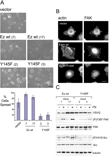
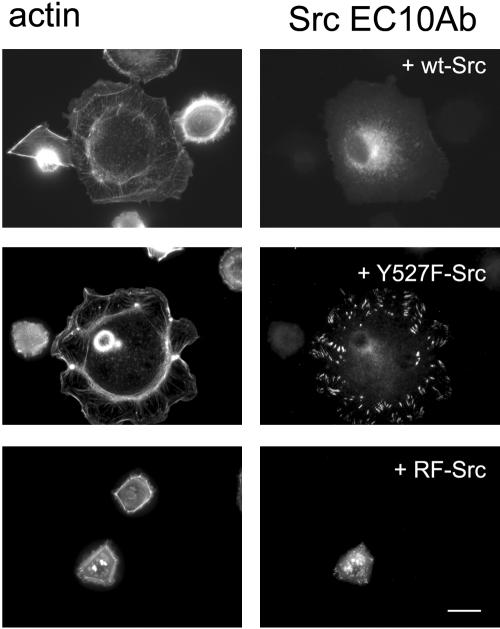
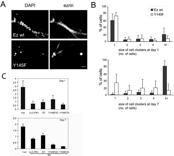
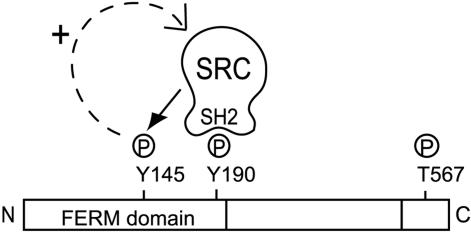
In addition to providing a regulated linkage between the membrane and the actin cytoskeleton, ezrin participates in signal transduction pathways. Here we describe that expression of the ezrin Y145F mutant delays epithelial cell spreading on fibronectin by inhibiting events leading to FAK activation. The defect in spreading was rescued by the overexpression of catalytically functional Src. We demonstrate that ezrin Y145 is phosphorylated in A431 cells stimulated with epidermal growth factor (EGF) and in v-Src-transformed cells. Moreover in cells devoid of Src, SYF-/- fibroblasts, ezrin Y145 phosphorylation could only be detected upon the introduction of an active form of Src. The phosphorylation of ezrin at Y145 required prior binding of the Src SH2 domain to ezrin. Our results further show that Src activity influences its binding to ezrin and a positive feedback mechanism for Src-mediated Y145 phosphorylation is implied. Interestingly, cells expressing ezrin Y145F did not proliferate when cultured in a 3D collagen gel. Collectively, our results demonstrate a key signaling input of Src-dependent ezrin phosphorylation in adhesion-mediated events in epithelial cells.


Figures

Figure 1.
Figure 1.
Ezrin Y145F mutant delays cell spreading onto fibronectin. (A) Phase contrast images of LLC-PK1 cells expressing vector alone (vector), ezrin wild-type expressing cells (two independent clones 7 and 17) and ezrin Y145F expressing cells (clones 2 and 3). Scale bar, 50 μm. Suspended cells were plated onto plastic dishes precoated with fibronectin (FN) and allowed to adhere for 1.5 h. Delayed cell spreading is observed with ezrin Y145F mutant expressing cells. For each cell clone, 10 random field views were analyzed for the mean percentage of cells spread shown in barchart; error bars, ±SD. (B) Fluorescence with anti-FAK antibody and phalloidin was performed in cells plated onto fibronectin-coated dishes for 1.5 h. Vector control cells and wild-type ezrin overexpressing cells show stress fibers and a disperse, punctate staining of FAK. In cells expressing ezrin Y145F, the assembly of F-actin is perturbed and FAK is concentrated to the periphery of the nonspread cells. Scale bar, 10 μm. (C) Cells extracts from two independent clones of each wild-type or Y145F ezrin expressing cells were prepared either from suspension cells or after plating onto fibronectin and analyzed by immunoblotting. FN-induced FAK Y397 phosphorylation is detected only in cells expressing wild-type ezrin. Lower phosphorylation level of Src Y418 in the ezrin Y145F expressing cells is detected compared with the wild-type ezrin expressing cells. Anti-tubulin antibody shows equal protein load. The results shown are representative of five independent experiments.
Figure 2.
Figure 2.
Src activity is essential for the spreading of LLC-PK1 cells on fibronectin. Cells expressing ezrin Y145F mutant (clone 2 shown) transfected with chicken Src (wt-Src), activated mutant of Src (Y527F-Src), or kinase dead mutant of Src (RF-Src). Cells were stained for F-actin and exogenous Src using an antiavian specific Src antibody (EC10 antibody). Overexpression of wild-type and activated Src but not kinase dead Src in ezrin Y145F cells promotes cell spreading and induces F-actin assembly. Bar, 10 μm.
Figure 3.
Figure 3.
Ezrin Y145 phosphorylation by Src. (A) Tyrosine phosphorylation ((P)Y) and phosphorylated Y145 ezrin ((P)Y145) were detected on immunoprecipitated VSVG-ezrin from LLC-PK1 cells untreated or treated with 0.1 mM pervanadate for 5 min (-/+ pvd). Pretreatment with the Src kinase inhibitor, PP2 (20 min; 10 μM) inhibits ezrin Y145 phosphorylation. (B) Endogenous ezrin immunoprecipitated from A431 cells. EGF treatment (3 min; 100 nM) induces ezrin Y145 phosphorylation, which is sensitive to the PP2 inhibitor. (C) Immunoprecipitated ezrin from MDCK ts v-Src cells at different times after v-Src activation (40.5°C switched to 35°C). Immunoblots detect time-dependent induced ezrin tyrosine phosphorylation and ezrin (P)Y145 upon v-Src activation. (D) Immunoprecipitated ezrin from SYF-/- cells. Only in cells transfected with Src Y527F is ezrin phosphorylated at Y145. (E) Src directly phosphorylates ezrin at Y145 in vitro. Top: kinase assays show phosphorylation of GST-truncated ezrin (Ez wt Δ52), but not of GST-wild-type ezrin (Ez wt). Reduced phosphorylation was detected on GST-truncated ezrin with the Y145F mutation. Bottom: Coomassie gel stains GST-purified ezrin variants used in the assay. Results are representative of three independent experiments. (F) VSVG-tag immunoprecipitations from cells expressing wild-type ezrin or the PIP2- mutant of ezrin. Pervanadate treatment does not induce Y145 phosphorylation of the ezrin PIP2- mutant.
Figure 4.
Figure 4.
Src SH2 domain binding to ezrin precedes Y145 phosphorylation. (A) GST or GST-Src SH2 domain were incubated with extracts of pervanadate-treated cells expressing ezrin wild-type and PIP2- ezrin. The proteins bound to the beads were subjected to SDS-PAGE and immunoblotted. Total cell lysate (VSVG input) and GST proteins (GST) were detected. Wild-type ezrin can interact with the SH2 domain of Src, whereas the PIP2- mutant of ezrin cannot. (B) The mutations Y190F and Y190F/Y240F abolish the interaction of ezrin with the SH2 domain of Src. (C) Reduced tyrosine phosphorylation, (P)Y, was observed on immunoprecipitated ezrin mutants Y145F and Y190F from pervanadate-treated cells compared with wild-type ezrin. Both ezrin Y145F and Y190F mutants showed a complete lack of immunoreactivity with the anti-(P)Y145 ezrin antibody. Results are representations of at least three experiments. (D) Cell extracts from wild-type or Y190F ezrin expressing cells were prepared either from suspension cells or after plating onto fibronectin (FN) and analyzed by immunoblotting. FN induced FAK Y397 phosphorylation is detected only in cells expressing wild-type ezrin. A lower phosphorylation level of Src Y418 is detected in cells expressing ezrin Y190F than in cells expressing wild-type ezrin.
Figure 5.
Figure 5.
Y145 phosphorylation sustains Src binding to ezrin. (A) GST or GST-Src SH2 domain were incubated with extracts of pervanadate-treated cells expressing ezrin wild-type and Y145F ezrin. Ezrin Y145F shows reduced binding to the SH2 domain of Src compared with ezrin wild-type. (B) GST-Src SH2 domain pull-down experiments were performed with MDCK ts v-Src cells at either the nonpermissive temperature (40.5°C; time 0) or at different times of induced v-Src expression upon switch to the permissive temperature (35°C). An interaction with the SH2 domain of Src and endogenous ezrin is only detected in cells induced to express v-Src.
Figure 6.
Figure 6.
Proliferation of ezrin Y145F expressing cells is blocked when cultured in 3D collagen gel. Single cells expressing wild-type ezrin (clone 7) or ezrin Y145F mutant (clone 3) were embedded in a 3D collagen gel and cultured for 7 d in the presence of HGF. (A) Fluorescence images at day 7 with DAPI and anti-ezrin antibody. Wild-type ezrin expressing cells form branching tubules with ezrin localized to the inner/apical pole of the tubules. The ezrin Y145F expressing cells are seen as either single cells or in small clusters, with intact nuclei as observed by DAPI staining. Bar, 10 μm. (B) Barcharts show the mean percentage of cells ± SD, from 15 field views, in different sizes of cell clusters at either day 1 or day 7. Ezrin wild-type expressing cells show a shift to increased cell cluster size after day 7, in contrast to Y145F ezrin expressing cells. (C) Apoptosis of untransfected cells (LLC-PK1) and cells expressing either wild-type ezrin (E7) or ezrin Y145F (clones 2 and 3) was measured at day 1 (top graph) and 7 (bottom graph) of culture into collagen gel. At day 7 the values were taken of the cell sample diluted 4 times (4×). (+ve) is a positive control for detection of apoptosis. Error bars, ±SD. Culture of ezrin Y145F expressing cells in collagen does not result in increased cell apoptosis.
Figure 7.
Figure 7.
Feedback loop model of Src-mediated ezrin Y145 phosphorylation. Binding of Src SH2 domain to the putative phosphorylated Y190 is required and precedes Y145 phosphorylation. Ezrin Y145 phosphorylation affects the activation status of Src. The Src/ezrin interaction, by being dependent on Src activity, is positively influenced by ezrin Y145 phosphorylation.

Similar articles

Cited by

References

    1. Algrain, M., Turunen, O., Vaheri, A., Louvard, D., and Arpin, M. (1993). Ezrin contains cytoskeleton and membrane binding domains accounting for its proposed role as a membrane-cytoskeletal linker. J. Cell Biol. 120, 129-139. - PMC - PubMed
    1. Autero, M., Heiska, L., Ronnstrand, L., Vaheri, A., Gahmberg, C. G., and Carpen, O. (2003). Ezrin is a substrate for Lck in T cells. FEBS Lett. 535, 82-86. - PubMed
    1. Barret, C., Roy, C., Montcourrier, P., Mangeat, P., and Niggli, V. (2000). Mutagenesis of the phosphatidylinositol 4,5-bisphosphate (PIP(2)) binding site in the NH(2)-terminal domain of ezrin correlates with its altered cellular distribution. J. Cell Biol. 151, 1067-1080. - PMC - PubMed
    1. Behrens, J., Vakaet, L., Friis, R., Winterhager, E., Van Roy, F., Mareel, M. M., and Birchmeier, W. (1993). Loss of epithelial differentiation and gain of invasiveness correlates with tyrosine phosphorylation of the E-cadherin/beta-catenin complex in cells transformed with a temperature-sensitive v-SRC gene. J. Cell Biol. 120, 757-766. - PMC - PubMed
    1. Berryman, M., Gary, R., and Bretscher, A. (1995). Ezrin oligomers are major cytoskeletal components of placental microvilli: a proposal for their involvement in cortical morphogenesis. J. Cell Biol. 131, 1231-1242. - PMC - PubMed

Publication types

MeSH terms

LinkOut - more resources