. 2005 Jan 12;25(2):308-17.
doi: 10.1523/JNEUROSCI.3967-04.2005.

The micromolar zinc-binding domain on the NMDA receptor subunit NR2B

Affiliations

The micromolar zinc-binding domain on the NMDA receptor subunit NR2B

Julie Rachline et al. J Neurosci. .

Abstract

Eukaryotic ionotropic glutamate receptor subunits possess a large N-terminal domain (NTD) distinct from the neighboring agonist-binding domain. In NMDA receptors, the NTDs of NR2A and NR2B form modulatory domains binding allosteric inhibitors. Despite a high sequence homology, these two domains have been shown to bind two ligands of strikingly different chemical nature. Whereas the NTD of NR2A binds zinc with high (nanomolar) affinity, the NTD of NR2B binds the synthetic neuroprotectant ifenprodil and its derivatives. Using both NTD-mutated/deleted receptors and isolated NTDs, we now show that the NTD of NR2B, in contrast to NR2C and NR2D, also binds zinc, but with a lower affinity. Furthermore, we present evidence that zinc and ifenprodil compete for an overlapping binding site. This modulatory binding site accounts for the submicromolar zinc inhibition of NR1/NR2B receptors. Given that zinc is accumulated and released at many glutamatergic synapses in the CNS, these findings suggest that zinc is the endogenous ligand of the NTD of both NR2A and NR2B, the two major NR2 subunits. Thus, NMDA receptors contain zinc sensors capable of detecting extracellular zinc over a wide concentration range depending on their NR2 subunit composition. The coexistence of subunit-specific zinc-binding sites of high (nanomolar) and low (micromolar) affinity on NMDA receptors raises the possibility that zinc exerts both a tonic and a phasic control of membrane excitability.


Figures

Figure 7.
Figure 7.
Zn protects the isolated NTD of NR2B against trypsinolysis. Isolated wild-type and mutated NTDs of NR2B were produced in E. coli, purified, and subjected to trypsin proteolysis with or without Zn or ifenprodil for various amounts of time (up to 10 min). NTDs were produced either as a full-length version (main band at ∼40 kDa) or as a C-terminal truncated (tr) version (main band at ∼30 kDa) (Perin-Dureau et al., 2002). Lane 0 corresponds to the protein solution just before trypsin addition. Ifenprodil was used at 100 μm, and Zn was used at 300 μm. A, Wild-type NTD is protected against trypsinolysis by ifenprodil and Zn. B, NTD mutation D101A abolishes protection against proteolysis by either ligand. C, NTD mutation H127A specifically abolishes Zn-induced protection, leaving ifenprodil-induced protection unaffected.
Figure 3.
Figure 3.
The NTD of NR2B controls Zn inhibition of NR1/NR2B receptors. Comparison of the Zn dose-response curves obtained on NR1/NR2 receptors containing either wild-type or NTD-deleted NR2 subunits is shown. Data were fitted with the Hill equation. Each data point is the mean value from at least three different oocytes. A, Wild-type (wt) and NTD-deleted NR2A-containing receptors. Currents were measured at +50 mV. The estimated values of Zn IC50 are 16 nm (wt) and 12 μm (ΔNTD). B, Wild-type and NTD-deleted NR2B-containing receptors. Currents were measured at +50 mV. The estimated values of Zn IC50 are 760 nm (wt) and 12 μm (ΔNTD). For this latter construct, data points obtained with Zn concentrations >100 μm were excluded from the fit because Zn, in this concentration range, produced an additional potentiating effect. This effect could be attributable to Zn acting on the NR2B-specific Mg/spermine potentiating site (Paoletti et al., 1995). C, Wild-type and NTD-deleted NR2D-containing receptors. Currents were measured at -60 mV (for NR2D-containing receptors, at this potential and in the tested Zn concentration range, there is no significant contribution of the Zn voltage-dependent block; see Materials and Methods). The estimated values of Zn IC50 are 8.4 μm (wt) and 12.4 μm (ΔNTD). D, Effects of NR2 NTD deletions and transplantation on Zn sensitivity. Zn IC50 values are displayed on a logarithmic scale. Deleting the NTD of NR2A or NR2B strongly affects Zn sensitivity (750-fold and 16-fold increase in Zn IC50 value, respectively), whereas deleting the NTD of NR2D produces very little effect (1.3-fold increase in Zn IC50 value). Transplanting the NTD of NR2B on the NTD-deleted NR2A subunit [chimera NR2A (NTD-2B)] confers a Zn sensitivity close to that of wild-type NR1/NR2B (IC50 value of 1.45 μm, maximal inhibition close to 1) (Paoletti et al., 2000). Note that all NR2 NTD-deleted receptors display a nearly identical sensitivity to Zn, indicating the presence of a remaining low-affinity, voltage-independent and NR2 NTD-independent Zn-binding site common to all receptor subtypes.
Figure 1.
Figure 1.
The Zn sensitivity of recombinant NMDA receptors is NR2 specific. Zn dose-response curves of diheteromeric NR1/NR2 receptors expressed in Xenopus oocytes are shown. Currents were elicited by saturating concentrations of glutamate and glycine (100 μm each) and measured at +50 mV, at which voltage-dependent Zn block is virtually absent. For NR1/NR2A receptors, tricine was used to buffer Zn. For other receptor subtypes, Zn was not buffered, and Zn concentrations were corrected for contaminating Zn (for NR1/NR2B receptors, very similar curves were obtained using tricine-buffered solutions; see Materials and Methods). The curves represent least-square fits to the data point with the Hill equation. The estimated values of IC50, maximal inhibition, and Hill coefficient are as follows: 16 nm, 0.77 and 0.9 for NR1/NR2A receptors; 760 nm, 1.0 and 0.9 for NR1/NR2B receptors; 18 μm, 0.93 and 0.9 for NR1/NR2C receptors; 9.2 μm, 1.0 and 1.2 for NR1/NR2D receptors. Each data point is the mean value from at least three different oocytes.
Figure 5.
Figure 5.
Determinants controlling NR2-specific inhibition of NMDA receptor activity by Zn and ifenprodil. Sequence alignment of all four NR2 subunit NTDs was adapted from Paoletti et al. (2000). Residues of NR2A controlling the high-affinity Zn inhibition are shown in red [T104, a newly identified residue, is highlighted as the mutation 2A-T104A increases the Zn IC50 value by >60-fold (n = 4)] (P. Paoletti, unpublished data). Residues of NR2B are color-coded as follows: red for those controlling the intermediate-affinity Zn inhibition, green for those controlling ifenprodil inhibition, and blue for those controlling both Zn and ifenprodil inhibitions (see Fig. 4). Mutations of these critical residues all result in a decrease of Zn and/or ifenprodil sensitivity, except for mutation V42A, which decreases ifenprodil sensitivity but increases Zn sensitivity (see Results). Also note that mutating 2B-Y175 not only affects Zn inhibition but also slightly decreases ifenprodil sensitivity (see Fig. 4). The β-strands and α-helices identified in the structure of the related bacterial protein LIVBP (PDB 2liv) are indicated on top of the alignment; boxes indicate regions (mostly loops) known to contact the ligand molecules in LIVBP-like domains (Paoletti et al., 2000; Perin-Dureau et al., 2002).
Figure 2.
Figure 2.
Differential effects of NR2 NTD deletions on Zn sensitivity. Each trace shows the inhibition by Zn of NMDA NR1/NR2 receptors containing either wild-type (left) or NTD-deleted (ΔNTD; right) NR2 subunits. Recordings were made at -60 mV. The bars above the current traces indicate the duration of agonist (agos) and Zn applications. Diagrams of wild-type and deleted NR2 constructs are shown at the top (S1 and S2, agonist binding domains; 1, 3, and 4, transmembrane segments; 2, pore loop). A, A concentration of 200 nm Zn (buffered with tricine) on wild-type and deleted NR1/NR2A receptors. B, A concentration of 1 μm Zn on wild-type and deleted NR1/NR2B receptors. C, A concentration of 20 μm Zn on wild-type and deleted NR1/NR2D receptors.
Figure 4.
Figure 4.
Identifying residues in the NTD of NR2B controlling Zn inhibition of NR1/NR2B receptors. A, Zn dose-response curves of NR1/NR2B receptors mutated at positions NR2B-V42, NR2B-D101, and NR2B-H127. Whereas mutations D101A and H127A decrease Zn sensitivity (Zn IC50 value of 5.7 and 4.9 μm, respectively), mutation V42A increases Zn sensitivity (Zn IC50 value of 0.17 μm). The dotted curves are the fits of the Zn dose-response curves of wild-type NR1/NR2B receptors (left dotted curve) and NR1/NR2B-ΔNTD receptors (right dotted curves) obtained in Figure 3. For NR1/NR2B-V42A receptors, tricine-buffered Zn solutions were used. Each data point is the mean value from at least three different oocytes. B, Comparison of the effects of NR2B NTD mutants on ifenprodil (left) and Zn (right) inhibitions quantified as the ratio of the mean Zn IC50 value for mutant receptors over that of wild-type NR1/NR2B receptors. Listed are all residues that have been identified as critical for ifenprodil (Perin-Dureau et al., 2002) and Zn (present study) inhibitions. Mutations that had no significant effect on Zn or ifenprodil inhibition had ratio values fixed to 1 (dotted lines). Note that some mutations affect either Zn inhibition only or ifenprodil inhibition only, whereas others affect both.
Figure 6.
Figure 6.
A competitive interaction between Zn and ifenprodil on NR1/NR2B receptors. Off-relaxation kinetics of Zn and ifenprodil applied separately or simultaneously during NMDA responses recorded at -60 mV on Xenopus oocytes expressing wild-type NR1/NR2B receptors are shown. In all experiments, ifenprodil was applied at 200 nm, a concentration close to the IC50 value. Zn was applied at two different saturating concentrations, 20 μm (∼25-fold IC50 value) or 300 μm (∼400-fold IC50 value). A1-A4, All traces are from a single cell. A1, A2, Zn (20 μm) washout kinetic is much faster than that of ifenprodil. A3, A4, Zn (20 μm) is applied on receptors first equilibrated with ifenprodil. After simultaneous removal of both antagonists, a fast and a slow component are seen. When Zn is coapplied for a short period (10 sec), the weight of the fast and the slow component is about equal. In contrast, when Zn is coapplied for a long period (8 min), the fast component clearly predominates, indicating that Zn has displaced ifenprodil molecules. B1, B2, All traces are from a single cell. Similar protocols as above, but with an increased Zn concentration (300 μm), are shown. B1, Fast washout kinetic of Zn applied alone. B2, When Zn is coapplied for a long period (8 min), removal of both antagonists reveals a (single) fast component of wash only, demonstrating that Zn ions have entirely replaced ifenprodil molecules. Inset, Off-relaxations shown on an expanded time scale and after current normalization. Exponential fits are superimposed (dashed white lines) with time constants of 4.1 sec for Zn alone, 59.6 sec for ifenprodil alone, and 3.4 sec for coapplication (8 min) of Zn and ifenprodil.
Figure 8.
Figure 8.
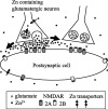
Dual modulation, tonic and phasic, of NMDA receptor activity by extracellular Zn. Zn is known to be concentrated at many glutamatergic synapses in the CNS. Both transport of Zn into neurons from the extracellular space and into synaptic vesicles have been characterized (Frederickson et al., 2000; Smart et al., 2004). In this model, we propose that ambient Zn levels exert a tonic inhibition of NMDA receptors through binding to the NR2A-specific nanomolar Zn-binding site. During synaptic activity, Zn is coreleased with glutamate in the synaptic cleft where Zn concentrations are expected to be high enough (micromolar range) to also inhibit the NR2B-specific micromolar Zn-binding site. Thick arrows indicate the possibility that, after phasic liberation, Zn diffuses outside the synaptic cleft. This spillover of Zn could induce inhibition of extrasynaptic NMDA receptors but also of NMDA receptors located at neighboring synapses.
None

References

    1. Armstrong N, Sun Y, Chen GQ, Gouaux E (1998) Structure of a glutamate-receptor ligand-binding core in complex with kainate. Nature 395: 913-917. - PubMed
    1. Ayalon G, Stern-Bach Y (2001) Functional assembly of AMPA and kainate receptors is mediated by several discrete protein-protein interactions. Neuron 31: 103-113. - PubMed
    1. Bogden JD, Troiano RA, Joselow MM (1977) Copper, zinc, magnesium, and calcium in plasma and cerebrospinal fluid of patients with neurological diseases. Clin Chem 23: 485-489. - PubMed
    1. Chen GQ, Cui C, Mayer ML, Gouaux E (1999) Functional characterization of a potassium-selective prokaryotic glutamate receptor. Nature 402: 817-821. - PubMed
    1. Chen N, Moshaver A, Raymond LA (1997) Differential sensitivity of recombinant N-methyl-d-aspartate receptor subtypes to zinc inhibition. Mol Pharmacol 51: 1015-1023. - PubMed

Publication types

LinkOut - more resources