. 2005 Jan 12;25(2):436-45.
doi: 10.1523/JNEUROSCI.1575-04.2005.

Longer forms of amyloid beta protein: implications for the mechanism of intramembrane cleavage by gamma-secretase

Affiliations

Longer forms of amyloid beta protein: implications for the mechanism of intramembrane cleavage by gamma-secretase

Yue Qi-Takahara et al. J Neurosci. .

Abstract

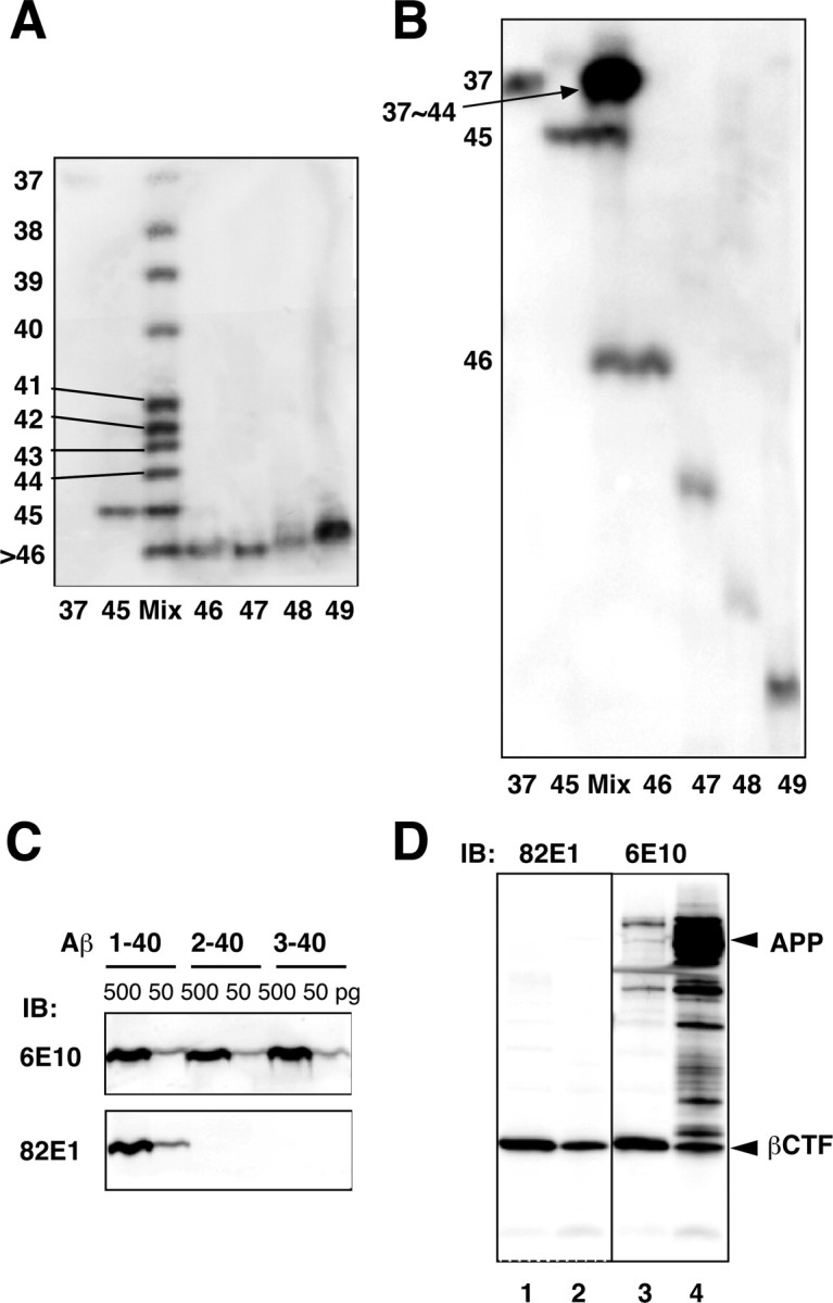
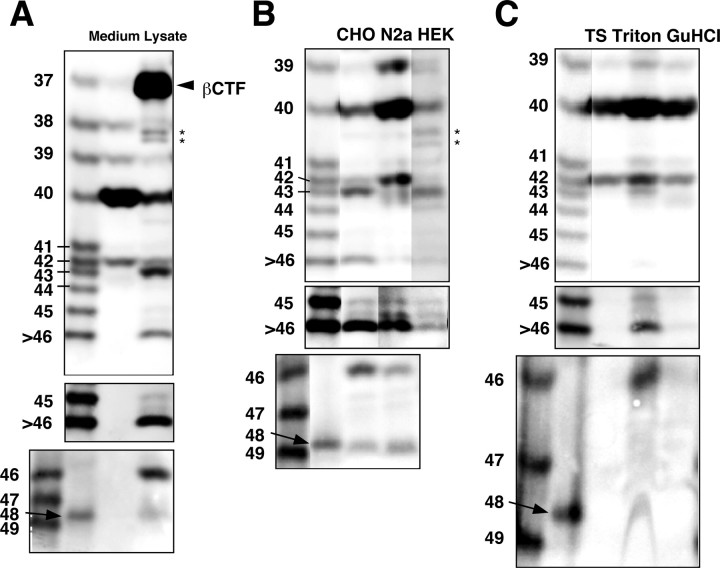
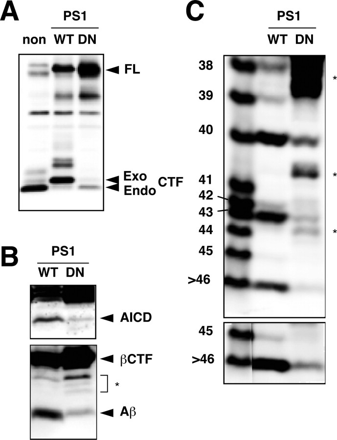
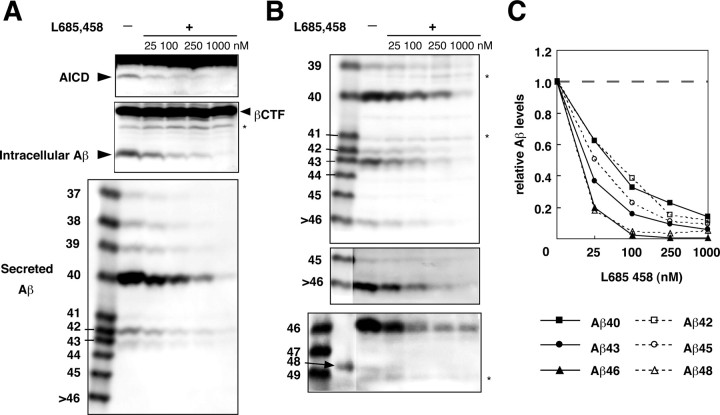
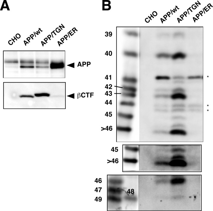
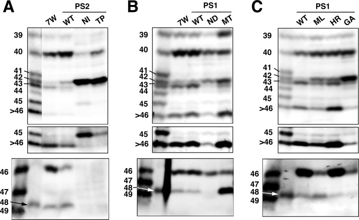
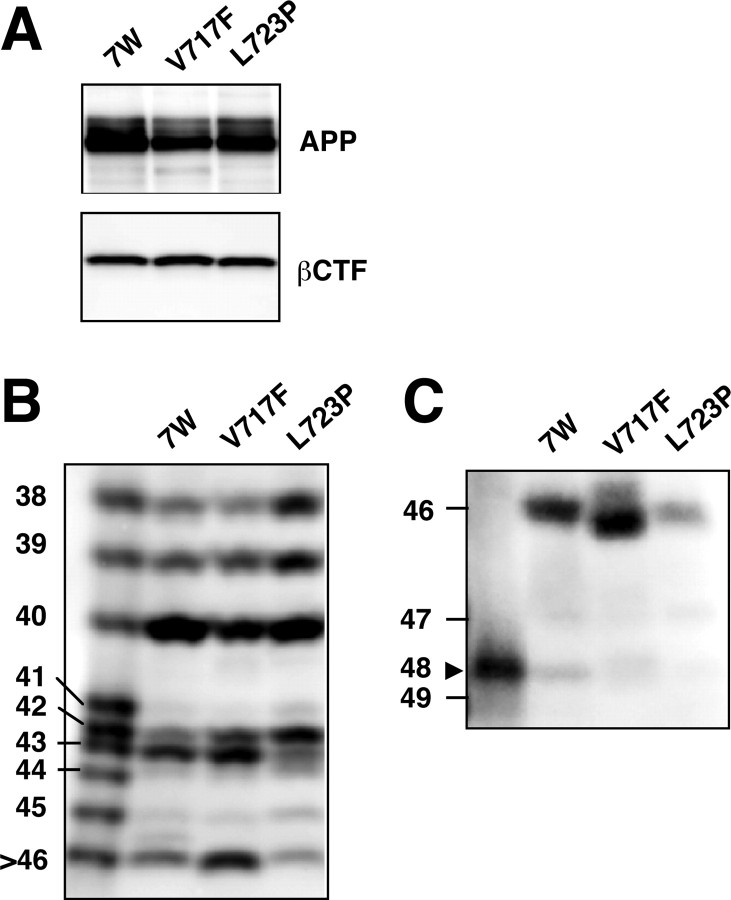
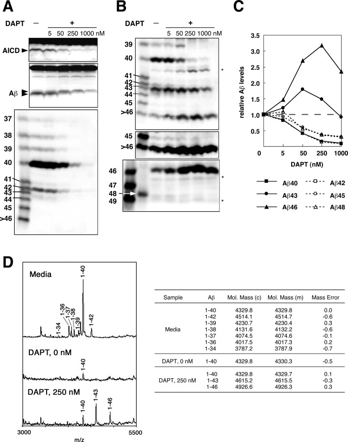
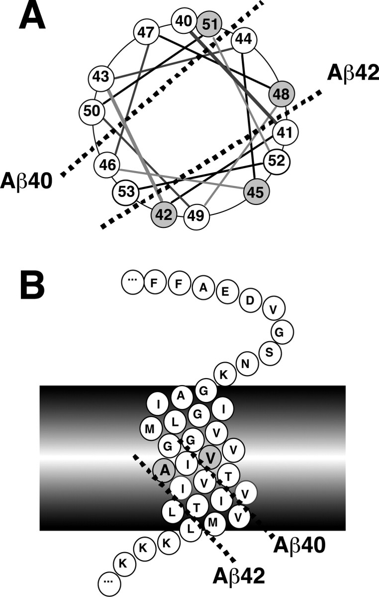
Gamma-cleavage of beta-amyloid precursor protein (APP) in the middle of the cell membrane generates amyloid beta protein (Abeta), and epsilon-cleavage, approximately 10 residues downstream of the gamma-cleavage site, releases the APP intracellular domain (AICD). A significant link between generation of Abeta and AICD and failure to detect AICD41-99 led us to hypothesize that epsilon-cleavage generates longer Abetas, which are then processed to Abeta40/42. Using newly developed gel systems and an N-end-specific monoclonal antibody, we have identified the longer Abetas (Abeta1-43, Abeta1-45, Abeta1-46, and Abeta1-48) within the cells and in brain tissues. The production of these longer Abetas as well as Abeta40/42 is presenilin dependent and is suppressed by {1S-benzyl-4R-[1S-carbamoyl-2-phenylethylcarbamoyl-1S-3-methylbutylcarbamoyl]-2R-hydroxy-5-phenylpentyl}carbamic acid tert-butyl ester, a transition state analog inhibitor for aspartyl protease. In contrast, N-[N-(3,5-difluorophenacetyl)-L-alanyl]-S-phenylglycine t-butyl ester, a potent dipeptide gamma-secretase inhibitor, builds up Abeta1-43 and Abeta1-46 intracellularly, which was also confirmed by mass spectrometry. Notably, suppression of Abeta40 appeared to lead to an increase in Abeta43, which in turn brings an increase in Abeta46, in a dose-dependent manner. We therefore propose an alpha-helical model in which longer Abeta species generated by epsilon-cleavage is cleaved at every three residues in its carboxyl portion.


Figures

Figure 1.
Figure 1.
Modified Tris/Tricine/8 m urea gels and specificity of 82E1. A, B, Synthetic Aβ1-37 through Aβ1-49 were separated using gel I (A) and gel II (B). In gel I (A), Aβ1-37 through Aβ1-45 are clearly separated, whereas Aβ1-46 through Aβ1-49 are stuck at the gel front. In gel II (B), Aβ1-45 through Aβ1-49 are well separated, whereas Aβ1-37 through Aβ1-44 are stuck at the upper region of the gel. C, Synthetic Aβ1-40, Aβ2-40, and Aβ3-40 (500 and 50 pg of each) were subjected to Western blotting using 6E10 (top) or 82E1 (bottom). Whereas 6E10 labeled all of these Aβs to similar extents, 82E1 recognized only Aβ1-40. D, The cell lysates from CHO cells overexpressing βCTF (lanes 1, 3) or APP (lanes 2, 4) were subjected to Western blotting with 82E1 (left) or 6E10 (right). 82E1 specifically labeled βCTF but not full-length APP or several APP-derived products that are strongly labeled with 6E10.
Figure 2.
Figure 2.
Longer Aβs in the various cell lysates and the APP-transgenic mouse brain. A, BAN50 immunoprecipitates from the conditioned medium (Medium) and Triton-soluble fraction (Lysate) of 7WD10 cells were separated on gel I (top and middle) and gel II (bottom) and subjected to Western blotting with 82E1. The middle panel represents an overexposed blot (the same as above). Aβ species longer than Aβ42 are undetectable in the conditioned medium but are detectable in the cell lysate. B, BAN50 immunoprecipitates of Triton-soluble fractions from CHO, N2a, and HEK cell lines stably expressing APP were separated on gel I (top and middle) and gel II (bottom) and subjected to Western blotting with 82E1. The middle panel represents an overexposed blot. Although the relative levels of Aβ species differ, the same longer Aβ species as found in CHO cells are detectable in the N2a and HEK cell lines. In N2a cell line, a band just above Aβ42 presumably represents Aβ41. The origins of an extra band below Aβ43 in the N2a lane and a band located between Aβ45 and Aβ46 seen in all three lanes are unknown. C, Aβ43, Aβ45, Aβ46, and Aβ48 are found exclusively in the Triton-soluble fraction of (plaque-free) brain homogenates from 2.5-month-old APP-transgenic mice (Tg2576). Immunoprecipitates from TBS- (TS), Triton-, and GuHCl-soluble fractions of Tg2576 mouse brain homogenates were separated on gel I (top and middle) and gel II ((TS) bottom) and subjected to Western blotting with 82E1. The middle panel represents an overexposed blot. A weakly immunoreactive band above Aβ42 in the Triton lane presumably represents Aβ41. The asterisks in A and B are presumably C-terminally truncated βCTFs. When different gel conditions are used, these bands exhibit the various mobilities relative to those of synthetic Aβs, which contrasts with the observation that longer Aβs identified here always comigrate with corresponding synthetic Aβs.
Figure 3.
Figure 3.
A DN mutant of PS1 greatly reduced intracellular levels of longer Aβs. A, CHO cells that inducibly express βCTF were stably transfected with cDNAs encoding wt (WT) or DN (D257A/D385A) mtPS1. Exogenous human wt or mtPS1 displaced endogenous PS1 to a large extent. Lysates were prepared from a nontransfectant (non) and the two transfectants, and equal amounts of protein were subjected to Western blotting with anti-G1L3. Full-length PS1 (FL) and endogenous (Endo) and exogenous (Exo) CTFs are indicated by arrowheads. B, The DN mtPS1 caused a remarkable reduction in the levels of AICD and Aβ in the lysate. After induction of βCTF for 4 hr, the lysates were prepared from these two transfectants, and equal amounts of protein were subjected to Western blotting using UT421 (top) or 82E1 (bottom). C, The levels of longer Aβs were also greatly suppressed by the expression of DN mtPS1. The immunoprecipitates from the Triton-soluble fraction of the two stable transfectants were separated on gel I and then subjected to Western blotting with 82E1. The bottom panel represents an overexposed blot. A couple of bands indicated by asterisks in B and C presumably represent C-terminally truncated βCTFs. The inhibition of γ-secretase caused an increase of those bands.
Figure 4.
Figure 4.
L-685,458 suppressed the levels of longer Aβs. A, The cells that inducibly express βCTF were first treated with indicated concentrations of L-685,458 for 2 hr, and βCTF was induced for 4 hr in the presence of L-685,458. Equal amounts of protein from whole-cell lysates were subjected to Western blotting with UT421 (top) and 82E1 (middle). The BAN50 immunoprecipitates from the medium were separated on gel I and subjected to Western blotting with 82E1 (bottom). B, Triton-soluble fractions of those treated cells were immunoprecipitated with BAN50, and the collected proteins were separated on gel I (top and middle) and gel II (bottom) and subjected to Western blotting with 82E1. The middle panel represents an overexposed blot. The asterisks in A and B indicate C-terminally truncated βCTFs. C, The amounts of intracellular Aβ were quantified using LAS-1000plus luminescent image analyzer. The levels of each Aβ species were normalized to those in the nontreated cells. The data shown are the means of the values from three (for Aβ40, Aβ42, Aβ43, and Aβ45) or two (for Aβ46 and Aβ48) independent experiments.
Figure 5.
Figure 5.
Longer Aβ forms are produced at the same location as Aβ40/42. A, Stable cell lines overexpressing wtAPP (APP/wt), APP carrying a TGN38 sorting signal (APP/TGN), or an ER retention signal (APP/ER) were generated. The cell lysates with the same amounts of protein were subjected to Western blotting with UT421 for full-length APP (top) or with 82E1 for βCTF (bottom). B, Triton-soluble fractions from these cell lines with the same amounts of protein were immunoprecipitated with BAN50. The immunoprecipitates were separated on gel I (top and middle) and gel II (bottom) and subjected to Western blotting with 82E1. The middle panel represents an overexposed blot. The longer Aβ forms, including Aβ43, Aβ45, Aβ46, and Aβ48, were clearly visible in the APP/TGN cells. A couple of bands indicated by asterisks in B are presumably C-terminally truncated βCTFs.
Figure 6.
Figure 6.
MtPS1/2 altered the intracellular levels of longer Aβs. A, Immunoprecipitates from the lysates of 7WD10 (7W) cells, wt (WT), N141I (NI), and T122P (TP) mtPS2 transfectants were separated on gel I (top and middle) and gel II (bottom). The middle panel represents an overexposed blot. Striking increases in the Aβ42 levels and concomitant decreases in the Aβ40 levels in N141I and T122P mtPS2 cells were noted. B, Similarly, immunoprecipitates from 7WD10 (7W) cells, wt (WT), N135D (ND), and M233T (MT) mtPS1 transfectants were separated on gel I (top and middle) and gel II (bottom). The middle panel represents an overexposed blot. N135D PS1 is homologous to N141I PS2 and showed similar alterations in the intracellular Aβ levels. A remarkable increase in the Aβ48 level was a characteristic of M233T mtPS1. C, Immunoprecipitates from wt (WT), M146L (ML), H163R (HR), and G384A (GA) mtPS1 transfectants were separated on gel I (top and middle) and gel II (bottom). A middle panel represents an overexposed blot. All of these blots were probed with 82E1.
Figure 7.
Figure 7.
MtAPPs also altered the intracellular levels of longer Aβ forms. A, The levels of APP and βCTF were similar among 7WD10, V717F, and L723P mtAPP cells. The cell lysates were subjected to Western blotting with UT421 (top) or 82E1 (bottom). B, C, The lysates of these cells were immunoprecipitated with BAN50, and the precipitated proteins were separated on gel I (B) and gel II (C) and subjected to Western blotting with 82E1. A slightly faster mobility of Aβ46 in the V717F lane probably reflects V/F substitution at the position of Aβ46.
Figure 8.
Figure 8.
Differential effects of DAPT on the levels of longer Aβs. A, The cells that inducibly express βCTF were treated with the indicated concentrations of DAPT for 2 hr, and βCTF was induced for 4 hr in the presence of DAPT. Equal amounts of protein from the cell lysates were subjected to conventional Tris/Tricine gel electrophoresis, followed by Western blotting with UT421 to detect AICD (top) and with 82E1 to detect intracellular Aβ (middle). The relatively broad band representing intracellular Aβ apparently consists of two components that have slow and fast mobilities, as indicated by arrowheads. The fast-migrating Aβ component declined at 5 nm DAPT and was not discernible at 50 nm, where as the slow-migrating Aβ component appeared to increase at 50 nm and was discernible even at 1000 nm. The conditioned media were immunoprecipitated with BAN50, separated on gel I, and subjected to Western blotting with 82E1 (bottom). B, The lysates from those DAPT-treated cells were immunoprecipitated with BAN50, separated on gel I (top and middle) or gel II (bottom), and subjected to Western blotting with 82E1. The middle panel represents an overexposure of the top blot. A couple of bands indicated by asterisks are presumably C-terminally truncated βCTFs. C, The amounts of intracellular Aβ were quantified using LAS-1000plus luminescent image analyzer. The levels of each Aβ species were normalized to those in the nontreated cells. The data shown are the means of the values from three (for Aβ40, Aβ42, Aβ43, and Aβ45) or two (for Aβ46 and Aβ48) independent experiments. D, Mass spectra of secreted Aβ (top left) and intracellular Aβ from cells treated without and with 250 nm DAPT (middle and bottom left, respectively) after immunoprecipitation using monoclonal antibodies 4G8 and 6E10. The calculated (c) and observed (m) masses are shown in the right panel. The suppression of secreted Aβs by DAPT was very similar to that by L-685,458. However, the effects on the intracellular Aβs were quite distinct between these two inhibitors (see Fig. 4).
Figure 9.
Figure 9.
An α-helical model showing processing of longer Aβs to Aβ40/42. A, A view from the luminal side on the α-helix wheel representing a carboxyl half of the transmembrane domain of APP. The number follows Aβ numbering. The cleavage sites for generation of Aβ40 and Aβ42 (indicated by broken lines) are topographically in the opposite directions relative to the α-helical surface of the transmembrane domain. The carboxyl sides of Val-46 and Thr-43 are aligned with that of Val-40 on the same side of the α-helical surface. In contrast, the carboxyl sides of Thr-48 and Ilu-45 are aligned with that of Ala-42 on the opposite side. B, A side view on the α-helix of the transmembrane domain of APP. The cleavage sites for generation of Aβ40 and Aβ42 are distinctly aligned (indicated by broken lines) on the surface of the α-helix of the transmembrane domain.

Similar articles

Cited by

References

    1. Beher D, Wrigley JD, Owens AP, Shearman MS (2002) Generation of C-terminally truncated amyloid-β peptides is dependent on γ-secretase activity. J Neurochem 82: 563-575. - PubMed
    1. Citron M, Oltersdorf T, Haass C, McConlogue L, Hung AY, Seubert P, VigoPelfrey C, Lieberburg I, Selkoe DJ (1992) Mutation of the β-amyloid precursor protein in familial Alzheimer's disease increases β-protein production. Nature 360: 672-674. - PubMed
    1. Clarke NJ, Tomlinson AJ, Ohyagi Y, Younkin S, Naylor S (1998) Detection and quantitation of cellularly derived amyloid β peptides by immunoprecipitation-HPLC-MS. FEBS Lett 430: 419-423. - PubMed
    1. De Strooper B, Saftig P, Craessaerts K, Vanderstichele H, Guhde G, Annaert W, Von Figura K, Van Leuven F (1998) Deficiency of presenilin-1 inhibits the normal cleavage of amyloid precursor protein. Nature 391: 387-390. - PubMed
    1. Dovey HF, John V, Anderson JP, Chen LZ, de Saint Andrieu P, Fang LY, Freedman SB, Folmer B, Goldbach E, Holsztynska EJ, Hu KL, Johnson-Wood KL, Kennedy SL, Kholodenko D, Knops JE, Latimer LH, Lee M, Liao Z, Lievergurg IM, Motter RN, et al. (2001) Functional γ-secretase inhibitors reduce β-amyloid peptide levels in brain. J Neurochem 76: 173-181. - PubMed

Publication types

MeSH terms

LinkOut - more resources