. 2005 Jan 12;25(2):479-87.
doi: 10.1523/JNEUROSCI.2699-04.2005.

Postsynaptic shank antagonizes dendrite branching induced by the leucine-rich repeat protein Densin-180

Affiliations

Postsynaptic shank antagonizes dendrite branching induced by the leucine-rich repeat protein Densin-180

Arne Quitsch et al. J Neurosci. .

Abstract

Leucine-rich repeat and PDZ [postsynaptic density-95 (PSD-95)/Discs large/zona occludens-1] domain proteins such as scribble and Densin-180 have been implicated in the establishment of cell-cell contacts. Here, we show that Densin-180, which has been identified as a constituent of the postsynaptic density in excitatory synapses interacts with the postsynaptic scaffold protein shank (shank1-3). The interaction involves a two-point attachment of the C-terminal region of Densin-180 with the Src homology 3 domain and the N-terminal part of the proline-rich region of shank proteins. The N-terminal leucine-rich repeat region, which is not involved in binding shank, targets Densin-180 to the plasma membrane in transfected cells and to the basolateral membrane of epithelial cells. Nevertheless, coexpression of shank leads to a redirection of Densin-180 into intracellular clusters. In cultured hippocampal neurons, Densin-180 overexpression induces excessive branching of neuronal dendrites, which occurs at the expense of clusters for the postsynaptic marker PSD-95. Coexpression of shank3 abrogates branch formation and targets Densin-180 into postsynaptic clusters instead. Shank blocks binding of delta-catenin but not alphaCaM kinase II to Densin-180; because delta-catenin has been shown to induce branching and neurite formation, our data suggest a mechanism where shank could block the activation of a Densin-180-dependent signaling pathway by delta-catenin.


Figures

Figure 1.
Figure 1.
Interaction of the SH3 domain of shank1 with Densin-180. A, Domain structure of Densin-180. The position of the yeast two-hybrid (YTH) clone obtained with the shank1 SH3 domain is indicated. LAPSD, LAP-specific domain; UTR, untranslated region. B, Mapping of the interaction in the yeast two-hybrid system. Shank and Densin-180 constructs in pAS-2 and pACT2 were cotransformed into yeast strain CG-1945; protein interaction, as detected by growth on His-deficient media, was scored as ++, +, or -. C, Mapping of the interaction in transfected HEK cells. A shank1 construct containing the SH3 and PDZ domains was transfected into HEK cells together with N-terminal Flag epitope-tagged Densin-180 constructs as indicated. From cell lysates in RIPA buffer, shank was precipitated using GKAP peptide Sepharose. Precipitates (P) were analyzed by Western blotting using anti-shank PDZ and anti-Flag (Densin-180) antibodies. In, Input.
Figure 2.
Figure 2.
Densin-180 binds to shank proteins via a second point of interaction in the proline-rich region of shank. A, Densin-180 constructs containing Flag (top 5 panels), EGFP (panel 6), or myc-tags (bottom 2 panels) were coexpressed with shank cDNA constructs or T7 epitope-tagged βPIX in HEK cells, as indicated. Shank was precipitated from cleared cell lysates using GKAP Sepharose; βPIX was precipitated using T7-antibody agarose. Precipitates (P) were analyzed by Western blotting for the presence of Densin-180 (left panels; using anti-Flag), shank (right, top 3 panels; using anti-shank-PDZ), or βPIX (bottom right; using anti-T7). B, Deletion mapping in transfected HEK cells. Shank2 (left) and Densin-180 constructs (right) were cotransfected in HEK cells as indicated. The shank2 domain structure containing PDZ, proline-rich (pro), and sterile α motifs (SAM) is indicated. After cell lysis and precipitation of shank using GKAP Sepharose, shank and Densin-180 were detected by Western blotting using anti-shank PDZ and anti-Flag antibodies, respectively. C, His6-tagged Densin-180 (residues 1242-1537) was incubated with glutathione beads carrying GST fusions of various parts of shank proteins, as indicated. After washing, bound proteins were eluted with SDS sample buffer and analyzed by Western blotting using a His6-tag antibody. In, Input.
Figure 3.
Figure 3.
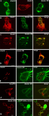
Shank targets Densin-180 into intracellular clusters. Expression vectors for EGFP-shank1 (A), Shank1-3 without tag (B-G), βPIX, EGFP-δ-catenin, and Densin-180 were transfected into HEK cells either alone (A) or in combination (B-G). Shank was visualized by staining using EGFP autofluorescence (A) or with rbAnti-shank, followed by Cy3-labeled anti-rabbit antibodies (red fluorescence). Densin-180 was visualized by monoclonal mouse anti-myc antibody, followed by Cy2-labeled anti-mouse antibodies (A-C, green fluorescence), by rbAnti-Densin-180, followed by Cy2-labeled anti-rabbit (D, red fluorescence), or by mouse anti-myc, followed by Cy3-labeled anti-mouse (E, red fluorescence). βPIX was labeled by anti-T7, followed by Cy3-labeled anti-mouse (D, red fluorescence), and δ-catenin was visualized by the EGFP-autofluorescence (E). All pictures are confocal sections taken approximately at the level of the center of the cells. Scale bars, 5 μm.
Figure 4.
Figure 4.
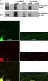
Shank and Densin-180 interact in vivo. A, Coprecipitation. Rat brain tissue was solubilized in deoxycholate buffer and cleared by centrifugation (Input). The lysate was subjected to immunoprecipitation, using rbAnti-Densin-180 or control antisera (left panels), or to peptide affinity purification, using GKAP C-terminal peptide, or a control peptide immobilized on NHS Sepharose (right panels). All samples were analyzed by Western blotting using shank-specific and Densin-180-specific antisera, as indicated. B, Colocalization. Hippocampal neurons were kept in culture for 17 d; after fixation with methanol, neurons were stained with affinity-purified gpAnti-shank, followed by Cy3-labeled anti-guinea pig secondary antibodies (middle panel) and affinity-purified rbAnti-Densin-180, followed by Alexa488-labeled anti-rabbit (top panel). Shown are overviews (left) and enlargements (right). Overlay of both fluorescence signals demonstrates extensive colocalization of both proteins in a punctate pattern (yellow staining; bottom panel). Scale bar, 5 μm.
Figure 5.
Figure 5.
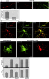
Densin-180 induces branching of neuronal dendrites. A, Hippocampal neurons were kept for 7 d in culture. After transfection with vectors coding for EGFP or EGFP-Densin-180, cells were cultivated for another 7 d (7 + 7) and stained for EGFP. B, Costaining for MAP2. Neurons were transfected after 7 d in culture with the Densin-180-myc construct; after 2 d (7 + 2) or 7 d (7 + 7) in culture, cells were fixed, and overexpressed Densin-180 was detected by anti-myc, followed by Cy3-anti-mouse. MAP2 was detected using a polyclonal MAP antiserum (kindly provided by Dr. Stefan Kindler, Institut für Zellbiochemie, Hamburg, Germany), followed by Cy2-anti rabbit. C, Mapping of functional domains of Densin-180. Neurons were transfected with various deletion constructs of Densin-180, as indicated (top panel) and stained for the presence of Densin-180 using the ant-myc antibody as before. In parallel, constructs were transfected into HEK293 and assayed for their ability to target the protein to the plasma membrane. Note that only constructs that contain the leucine-rich repeat region are localized at the plasma membrane in HEK293 cells and have the ability to induce branching of dendrites in transfected neurons. Scale bars: neurons, full view, 20 μm; dendrite enlargements, HEK cells, 5 μm. D, MDCK-II cells stably expressing the Densin-180 LRR region (Densin1-470) were stained for Densin (green) and the tight junction marker ZO-1 (red; vertical sections only). Horizontal xy sections (left) were recorded on the confocal microscope; vertical xz sections (right) were then reconstructed using LSM software. Note the delineation of green (Densin) fluorescence by the red (ZO-1) signal. Scale bar, 5 μm; thickness of xz sections, 7 μm.
Figure 6.
Figure 6.
Relation of dendrite branching to the formation of postsynaptic complexes. A, Dendrites of control neurons (left) or neurons overexpressing Densin-180 (middle, right) were stained for the postsynaptic marker protein PSD-95 (left, middle) or the overexpressed Densin-180 (right). The number of PSD-95 puncta per 50 μm sections of individual dendrites was evaluated; *** significantly different from Densin-180-expressing cells, p < 0.0001, as determined by two-tailed Student's t tests. Scale bar, 5 μm. B, After 7 d in culture, neurons were cotransfected with constructs coding for EGFP-Densin-180 and myc-tagged Shank3/ProSAP2 (top panel), the leucine-rich region of Densin-180 (Densin-LRR) alone (second panel, left), or in combination with shank3 (second panel, middle and right). After 7 additional days, neurons were fixed and stained for Shank3 and EGFP-Densin-180. Note the extensive, punctate colocalization of both proteins in the merged picture for full-length Densin-180 (and Shank3). Transfected cells from this figure and Figure 5A were evaluated for their TDBTN and the number of branch points (>35 neurons for each experimental condition; *** significantly different from Densin-180-expressing cells; p < 0.0001), as determined by two-tailed Student's t tests. Scale bar, 20 μm; inset, 5 μm.
Figure 7.
Figure 7.
Shank blocks the interaction of the δ-catenin C terminus with the PDZ domain of Densin-180. A, B, HEK cells were cotransfected with the expression vectors indicated. After cell lysis, shank was precipitated using GKAP Sepharose; precipitates were analyzed by Western blotting. Note that δ-catenin, but not CaMKIIα, is excluded from the Densin-180/shank complex. C, HEK cells were transfected and lysed as above; Densin-180 was precipitated using the C-terminal peptide derived from δ-catenin, immobilized on NHS-Sepharose. Precipitates were analyzed by Western blotting. In, Input; P, precipitate.

Similar articles

Cited by

References

    1. Apperson ML, Moon IS, Kennedy MB (1996) Characterization of Densin-180, a new brain-specific synaptic protein of the O-sialoglycoprotein family. J Neurosci 16: 6839-6852. - PMC - PubMed
    1. Bilder D, Perrimon N (2000) Localization of apical epithelial determinants by the basolateral PDZ protein Scribble. Nature 403: 676-680. - PubMed
    1. Bilder D, Schober M, Perrimon N (2003) Integrated activity of PDZ protein complexes regulates epithelial polarity. Nat Cell Biol 5: 53-58. - PubMed
    1. Blichenberg A, Schwanke B, Rehbein M, Garner CC, Richter D, Kindler S (1999) Identification of a cis-acting dendritic targeting element in MAP2 mRNAs. J Neurosci 19: 8818-8829. - PMC - PubMed
    1. Böckers TM, Kreutz MR, Winter C, Zuschratter W, Smalla KH, Sanmarti-Vila L, Wex H, Langnaese K, Bockmann J, Garner CC, Gundelfinger ED (1999) Proline-rich synapse-associated protein-1/cortactin binding protein 1 (ProSAP1/CortBP1) is a PDZ-domain protein highly enriched in the postsynaptic density. J Neurosci 19: 6506-6518. - PMC - PubMed

Publication types

MeSH terms

LinkOut - more resources