

Proteomic analysis of mantle-cell lymphoma by protein microarray
- PMID: 15650054
- PMCID: PMC1895014
- DOI: 10.1182/blood-2004-10-3999
Proteomic analysis of mantle-cell lymphoma by protein microarray
Abstract
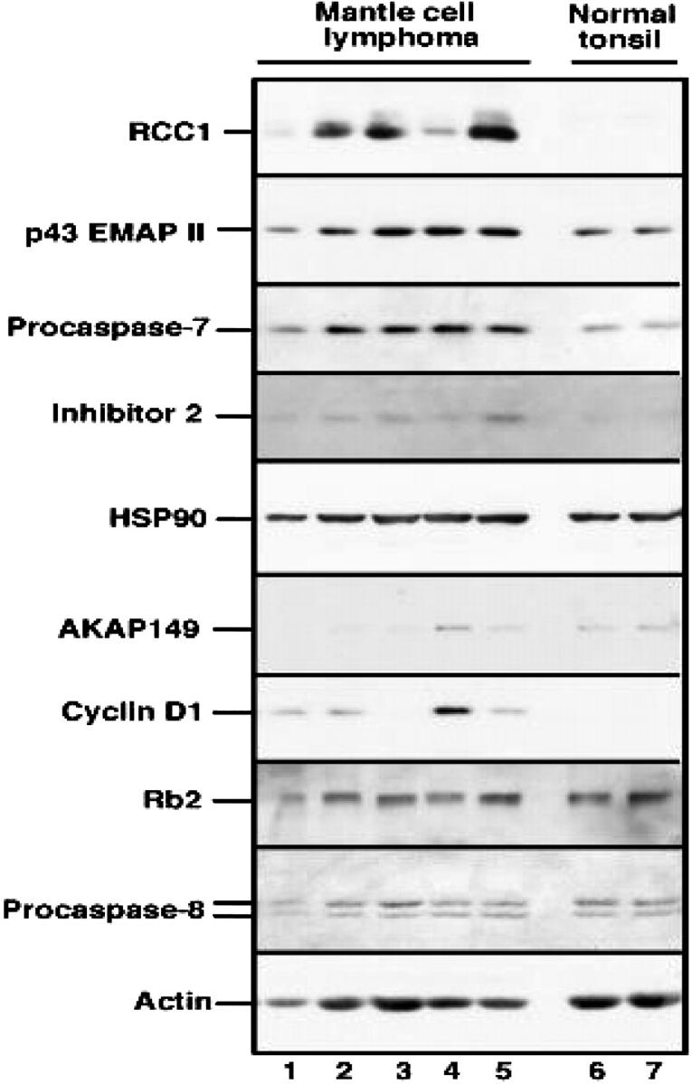
Mantle-cell lymphoma (MCL) is a unique subtype of B-cell non-Hodgkin lymphoma (NHL) that behaves aggressively and remains incurable. In order to understand the pathogenesis of MCL and design new therapies, it is important to accurately analyze molecular changes in pathways dysregulated in MCL. We used antibody microarrays to compare patterns of protein expression between CD19(+) purified B lymphocytes from normal tonsil and 7 cases of histologically confirmed MCL. Protein overexpression was defined as a higher than 1.3-fold or 2-fold increase in at least 67% of tumor samples compared with normal B-cell control. Of the polypeptides, 77 were overexpressed using the higher than 1.3-fold cutoff, and 13 were overexpressed using the 2-fold cutoff. These included cell cycle regulators (regulator of chromosome condensation 1 [RCC1], murine double minute 2 [MDM2]), a kinase (citron Rho-interacting kinase [CRIK]), chaperone proteins (heat shock 90-kDa protein [Hsp90], Hsp10), and phosphatase regulators (A-kinase anchor protein 1 [AKAP149], protein phosphatase 5 [PP5], and inhibitor 2). The elevated expression of some of these polypeptides was confirmed by immunoblotting and immunohistochemistry, whereas elevated expression of others could not be confirmed, illustrating the importance of confirmatory studies. This study describes a novel technique that identifies proteins dysregulated in MCL.
Figures


Similar articles
-
Protein profiling of plasma membranes defines aberrant signaling pathways in mantle cell lymphoma.Mol Cell Proteomics. 2009 Jul;8(7):1501-15. doi: 10.1074/mcp.M800515-MCP200. Epub 2009 Apr 2. Mol Cell Proteomics. 2009. PMID: 19346216 Free PMC article.
-
Two-dimensional molecular profiling of mantle cell lymphoma.Electrophoresis. 2003 Jul;24(14):2376-85. doi: 10.1002/elps.200305457. Electrophoresis. 2003. PMID: 12874873
-
Accuracy of diagnosing mantle cell lymphoma and identifying its variants on fine-needle aspiration biopsy.Cancer Cytopathol. 2019 Feb;127(1):44-51. doi: 10.1002/cncy.22077. Epub 2018 Nov 19. Cancer Cytopathol. 2019. PMID: 30452126
-
Altered apoptosis pathways in mantle cell lymphoma.Leuk Lymphoma. 2004 Jan;45(1):49-54. doi: 10.1080/1042819031000151112. Leuk Lymphoma. 2004. PMID: 15061196 Review.
-
Mantle cell lymphoma: 2015 update on diagnosis, risk-stratification, and clinical management.Am J Hematol. 2015 Aug;90(8):739-45. doi: 10.1002/ajh.24094. Am J Hematol. 2015. PMID: 26103436 Review.
Cited by
-
EMAP II Expression Is Increased on Peripheral Blood Cells from Non-Hodgkin Lymphoma.J Immunol Res. 2022 Sep 12;2022:7219207. doi: 10.1155/2022/7219207. eCollection 2022. J Immunol Res. 2022. PMID: 36132984 Free PMC article.
-
Transcriptional profiling of Epstein-Barr virus (EBV) genes and host cellular genes in nasal NK/T-cell lymphoma and chronic active EBV infection.Br J Cancer. 2006 Feb 27;94(4):599-608. doi: 10.1038/sj.bjc.6602968. Br J Cancer. 2006. PMID: 16449999 Free PMC article.
-
Identification of PGAM1 as a putative therapeutic target for pancreatic ductal adenocarcinoma metastasis using quantitative proteomics.Onco Targets Ther. 2018 Jun 6;11:3345-3357. doi: 10.2147/OTT.S162470. eCollection 2018. Onco Targets Ther. 2018. PMID: 29922073 Free PMC article.
-
Genetic heterogeneity and prognostic impact of recurrent ANK2 and TP53 mutations in mantle cell lymphoma: a multi-centre cohort study.Sci Rep. 2020 Aug 7;10(1):13359. doi: 10.1038/s41598-020-70310-9. Sci Rep. 2020. PMID: 32770099 Free PMC article.
-
Phosphorylation of RCC1 on Serine 11 Facilitates G1/S Transition in HPV E7-Expressing Cells.Biomolecules. 2021 Jul 6;11(7):995. doi: 10.3390/biom11070995. Biomolecules. 2021. PMID: 34356619 Free PMC article.
References
-
- Argatoff LH, Connors JM, Klasa RJ, Horsman DE, Gascoyne RD. Mantle cell lymphoma: a clinicopathologic study of 80 cases. Blood. 1997;89: 2067-2078. - PubMed
-
- Kurtin, PM. Mantle cell lymphoma. Adv Anat Pathol. 1998;5: 376-398. - PubMed
-
- Yatabe Y, Suzuki R, Matsuno Y, et al. Morphological spectrum of cyclin D1-positive mantle cell lymphoma: study of 168 cases. Pathol Int. 2001;51: 747-761. - PubMed
-
- Katz RL Wojcik EM, el-Naggar AK, Ordonez NG, Johnston DA. Proliferation markers in non-Hodgkin's lymphoma: a comparative study between cytophotometric quantitation of Ki-67 and flow cytometric proliferation index on fine needle aspirates. Anal Quant Cytol Histol. 1993;15: 179-186. - PubMed
-
- Weisenburger D, Vose JM, Greiner TC, et al. Mantle cell lymphoma: a clinicopathologic study of 68 cases from the Nebraska Lymphoma Study Group. Amer J Hematol. 2000;64: 190-196. - PubMed
Publication types
MeSH terms
Substances
Grants and funding
LinkOut - more resources
Full Text Sources
Other Literature Sources
Research Materials
