

Identification of a human B-cell/myeloid common progenitor by the absence of CXCR4
- PMID: 15650059
- PMCID: PMC1895023
- DOI: 10.1182/blood-2004-07-2839
Identification of a human B-cell/myeloid common progenitor by the absence of CXCR4
Abstract
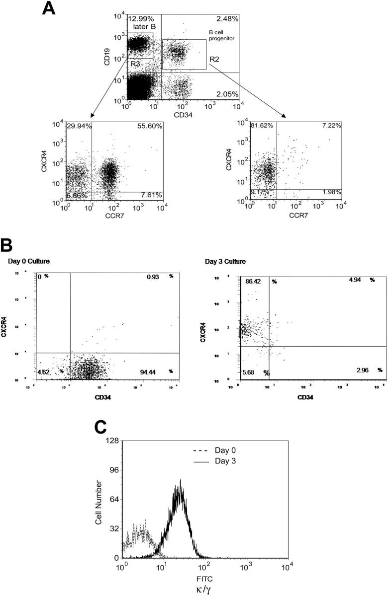
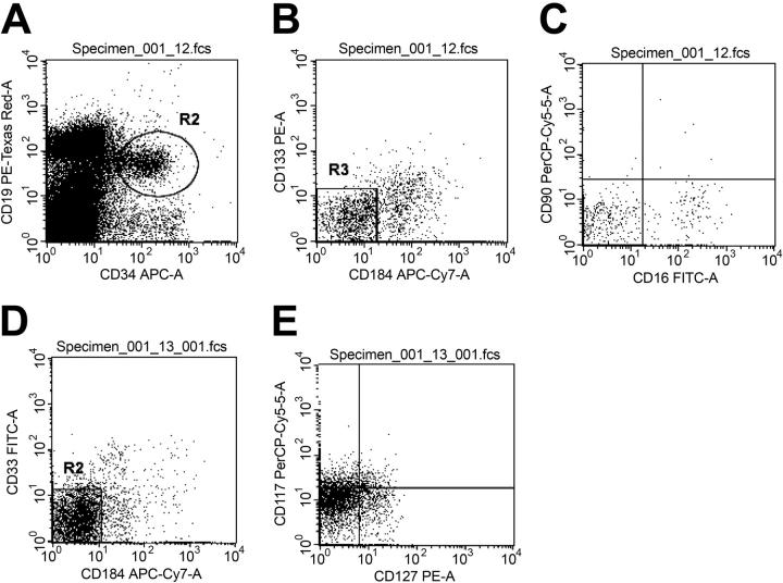
CXCR4 is a chemokine receptor required for hematopoietic stem cell engraftment and B-cell development. This study found that a small fraction of primitive CD34(+)/CD19(+) B-cell progenitors do not express CXCR4. These CD34(+)/CD19(+)/CXCR4(-) cells were also remarkable for the relative lack of primitive myeloid or lymphoid surface markers. When placed in B-lymphocyte culture conditions these cells matured to express CXCR4 and other surface antigens characteristic of B cells. Surprisingly, when placed in a myeloid culture environment, the CXCR4(-) B-cell progenitors could differentiate into granulocyte, macrophage, and erythroid cells at a high frequency. These data define a novel B-cell/myeloid common progenitor (termed the BMP) and imply a less restrictive pathway of myeloid versus lymphoid development than previously postulated.
Figures
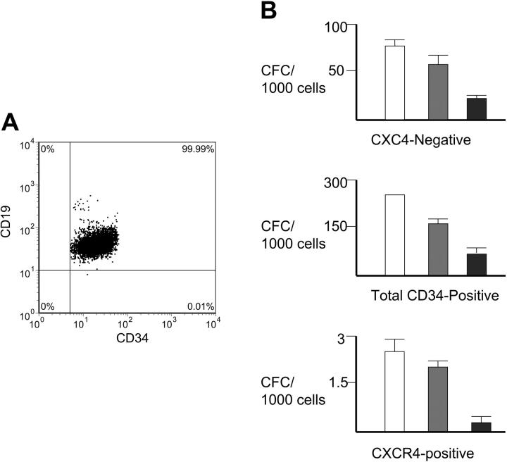
 , BFU-E; and ▪, CFU-GEMM. Note the different scales showing the colony numbers between the graphs; n = 3 each performed in triplicate. Error bars indicate standard error (SE).
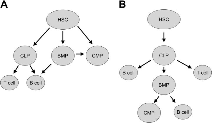
Similar articles
-
Expression of stromal cell-derived factor-1/pre-B cell growth-stimulating factor receptor, CXC chemokine receptor 4, on CD34+ human bone marrow cells is a phenotypic alteration for committed lymphoid progenitors.J Immunol. 1999 Oct 1;163(7):3612-20. J Immunol. 1999. PMID: 10490954
-
CXCL12-CXCR4 Axis Is Required for Contact-Mediated Human B Lymphoid and Plasmacytoid Dendritic Cell Differentiation but Not T Lymphoid Generation.J Immunol. 2017 Oct 1;199(7):2343-2355. doi: 10.4049/jimmunol.1700054. Epub 2017 Aug 25. J Immunol. 2017. PMID: 28842468
-
The heparin binding PECAM-1 adhesion molecule is expressed by CD34+ hematopoietic precursor cells with early myeloid and B-lymphoid cell phenotypes.Blood. 1993 Nov 1;82(9):2649-63. Blood. 1993. PMID: 7693043
-
Individual CD34+CD38lowCD19-CD10- progenitor cells from human cord blood generate B lymphocytes and granulocytes.Blood. 1997 May 15;89(10):3554-64. Blood. 1997. PMID: 9160660
-
Characterization and functional analysis of adult human bone marrow cell subsets in relation to B-lymphoid development.Blood. 1993 Jul 15;82(2):417-29. Blood. 1993. PMID: 7687158
Cited by
-
The phosphatidylserine receptors, T cell immunoglobulin mucin proteins 3 and 4, are markers of histiocytic sarcoma and other histiocytic and dendritic cell neoplasms.Hum Pathol. 2010 Oct;41(10):1486-94. doi: 10.1016/j.humpath.2010.04.005. Epub 2010 Jul 24. Hum Pathol. 2010. PMID: 20656318 Free PMC article.
-
KRAS mutation in secondary malignant histiocytosis arising from low grade follicular lymphoma.Diagn Pathol. 2018 Oct 15;13(1):78. doi: 10.1186/s13000-018-0758-0. Diagn Pathol. 2018. PMID: 30322385 Free PMC article.
-
CD34-positive cells and their subpopulations characterized by flow cytometry analyses on the bone marrow of healthy allogenic donors.Sao Paulo Med J. 2009 Jan;127(1):12-8. doi: 10.1590/s1516-31802009000100004. Sao Paulo Med J. 2009. PMID: 19466289 Free PMC article.
-
Differentiation Epitopes Define Hematopoietic Stem Cells and Change with Cell Cycle Passage.Stem Cell Rev Rep. 2022 Oct;18(7):2351-2364. doi: 10.1007/s12015-022-10374-4. Epub 2022 May 3. Stem Cell Rev Rep. 2022. PMID: 35503199 Free PMC article.
-
[Lineage switch from B cell to myeloid cell in the course of lymphoma treatment: three cases and literature review].Zhonghua Xue Ye Xue Za Zhi. 2018 Jun 14;39(6):518-520. doi: 10.3760/cma.j.issn.0253-2727.2018.06.017. Zhonghua Xue Ye Xue Za Zhi. 2018. PMID: 30032573 Free PMC article. Chinese. No abstract available.
References
-
- Bonnet D. Haematopoietic stem cells. J Pathol. 2002;197: 430-440. - PubMed
-
- Akashi K, Reya T, Dalma-Weiszhausz D, Weissman IL. Lymphoid precursors. Curr Opin Immunol. 2000;12: 144-150. - PubMed
-
- Kondo M, Scherer DC, Miyamoto T, et al. Cellfate conversion of lymphoid-committed progenitors by instructive actions of cytokines. Nature. 2000;407: 383-386. - PubMed
-
- Xie H, Ye M, Feng R, Graf T. Stepwise reprogramming of B cells into macrophages. Cell. 2004;117: 663-676. - PubMed
Publication types
MeSH terms
Substances
Grants and funding
LinkOut - more resources
Full Text Sources
Medical
