. 2005 Feb;79(3):1487-99.
doi: 10.1128/JVI.79.3.1487-1499.2005.

Sindbis virus translation is inhibited by a PKR/RNase L-independent effector induced by alpha/beta interferon priming of dendritic cells

Affiliations

Sindbis virus translation is inhibited by a PKR/RNase L-independent effector induced by alpha/beta interferon priming of dendritic cells

K D Ryman et al. J Virol. 2005 Feb.

Abstract

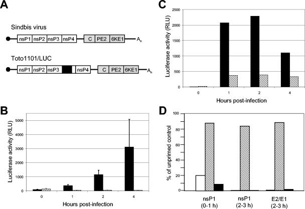
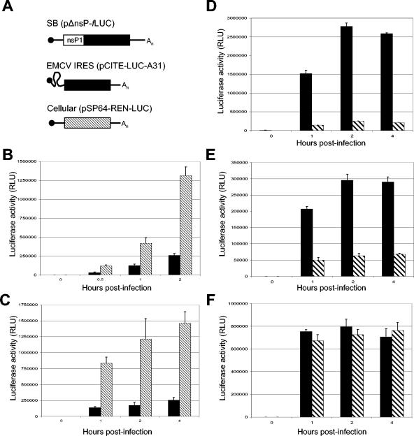
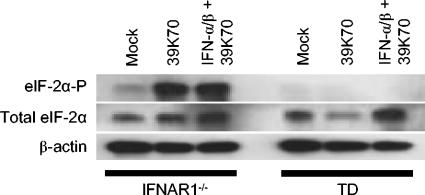
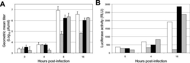
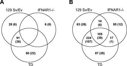
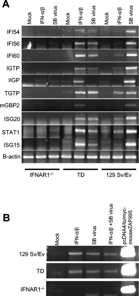
The tropism of Sindbis virus (SB) for cells of the dendritic cell (DC) lineage and the virulence of SB in vivo are largely determined by the efficacy of alpha/beta interferon (IFN-alpha/beta)-mediated antiviral responses. These responses are essentially intact in the absence of PKR and/or RNase L (K. D. Ryman, L. J. White, R. E. Johnston, and W. B. Klimstra, Viral Immunol. 15:53-76, 2002). In the present studies, we investigated the nature of antiviral effects and identity of antiviral effectors primed by IFN-alpha/beta treatment of bone marrow-derived DCs (BMDCs) generated from mice deficient in PKR and RNase L (TD). IFN-alpha/beta priming exerted significant antiviral activity at very early stages of SB replication and most likely inhibited the initial translation of infecting genomes. The early effect targeted cap-dependent translation as protein synthesis from an SB-like and a simple RNA were inhibited by interferon treatment, but an encephalomyocarditis virus internal ribosome entry site-driven element exhibited no inhibition. Phosphorylation of the alpha subunit of eukaryotic translation initiation factor 2 was defective after virus infection of TD cells, suggesting other mechanisms of translation inhibition. To identify components of these alternative antiviral pathway(s), we have compared global gene regulation in BMDCs derived from normal 129 Sv/Ev, IFNAR1-/-, and TD mice following infection with SB or treatment with IFN-alpha/beta. Candidate effectors of alternative antiviral pathways were those genes induced by virus infection or IFN-alpha/beta treatment in 129 Sv/Ev and TD-derived BMDC but not in virus-infected or IFN-alpha/beta-treated IFNAR1-/- cells. Statistical analyses of gene array data identified 44 genes that met these criteria which are discussed.


Figures

FIG. 1.
FIG. 1.
(A) Schematic representations of the genomes of unmodified SB and firefly luciferase (fLUC)-expressing Toto1101-LUC virus. NTRs (solid lines), nsP genes (open boxes), structural genes (shaded boxes), 7methyl-GTP cap (filled circle), poly(A) tail (An), and the fLUC ORF (black box within nsP3) are shown. (B) TD BMDC, either untreated (black bars) or primed for 6 h with 1,000 IU of IFN-α/β/ml (hatched bars), were infected with Toto1101/LUC (MOI = 10). At indicated times p.i., cells were lysed and luciferase activity was measured in relative light units (RLU). Data are means (n = 3), with error bars indicating standard deviations. Results are representative of two independent experiments. (C) Untreated (black bars) or IFN-α/β-primed (hatched bars) TD BMDC were harvested by using enzyme-free cell dissociation buffer and electroporated with capped Toto1101-LUC RNA transcripts. Luciferase activity was measured as described for panel B. (D) BMDC, either untreated or IFN-α/β primed, were infected with unmodified SB and pulsed with radiolabeled Cys and Met. Virus proteins were immunoprecipitated, separated by sodium dodecyl sulfate-PAGE, and quantitated on a phosphorimager. The accumulation of nsPs and E2/E1 in IFN-α/β-primed 129 Sv/Ev (white bars), IFNAR1−/− (hatched bars), and TD (black bars) BMDC are presented as a percentage of the untreated control cells. The time of the pulse is indicated.
FIG. 2.
FIG. 2.
Initial translation of virus genomes is suppressed in the absence of PKR. (A) Schematic representations of translational reporter constructs for SB (pSBΔnsP/fLUC), EMCV (pIRES-EMCV-A31), and host cell (pSP64-REN-LUC) mRNAs. (B and C) Capped SB (B) and EMCV IRES (C) translation reporter constructs (black bars) expressing fLUC were coelectroporated into BHK-21 cells with equimolar amounts of capped control RNA encoding rLUC (hatched bars). Cells were lysed at the indicated times, and rLUC and fLUC activities were measured with the dual-luciferase reporter assay system. Data are means (n = 3), with standard deviations indicated by error bars. (D to F) Untreated (black bars) or IFN-α/β-primed (hatched bars) TD BMDC were electroporated with capped SB fLUC-expressing translation reporter RNA transcripts (D), capped control RNA encoding rLUC (E), or EMCV IRES fLUC-expressing translation reporter RNA transcripts (F). Cells were lysed at the indicated times, and rLUC and fLUC luciferase activities are shown as means (n = 3) ± standard deviations.
FIG. 3.
FIG. 3.
eIF-2α phosphorylation in BMDC. IFNAR1−/− (lanes 1 to 3) and TD (lanes 4 to 6) BMDC were left untreated (lanes 1 and 4), infected with SB virus 39K70 (MOI = 10) for 12 h (lanes 2 and 5), or primed with 1,000 IU of IFN-α/β/ml for 6 h prior to infection (lanes 3 and 6). Levels of phosphorylated eIF-2α (eIF2α-P, top panel) and total eIF-2α (middle panel) were compared to levels of β-actin control (bottom panel) in cell extracts by immunoblot analysis. The results shown are representative of three independent experiments.
FIG. 4.
FIG. 4.
Priming is eliminated by ActD treatment. (A and B) Prior to virus infection, TD BMDC were untreated (white bars), primed with 1,000 IU of IFN-α/β for 6 h (hatched bars), treated with 1.0 μg of ActD/ml for 6 h (black bars), or ActD-treated for 1 h before priming for a further 5 h (shaded bars). (A) Cells were infected (MOI = 10) with unmodified SB virus, and at indicated times after infection, the accumulation of virus released into the cell supernatant was titrated by BHK-21 plaque assay. Data are presented as geometric means (n = 3) of log10 titers (PFU/ml) ± standard deviations. The experiment is representative of two independent experiments. (B) Cells were infected (MOI = 10) with fLUC-expressing Toto1101-LUC virus. Cells were lysed at the indicated times, and fLUC luciferase activities are shown.
FIG. 5.
FIG. 5.
Genes similarly and differentially induced by IFN-α/β priming and/or virus infection. Total RNA was isolated from 129 Sv/Ev, TD, and IFNAR1−/− BMDC either untreated, IFN-α/β primed (1,000 IU/ml for 6 h), or SB infected (12 h p.i.) and hybridized to murine U74Av2 GeneChip arrays (Affymetrix) with 12,488 gene expressions in each sample. Expression data were analyzed as described in Materials and Methods. Spotfire software was used to generate lists of genes that were upregulated ≥2- or ≥4-fold over untreated controls in replicate analyses (n = 2). Venn diagrams were generated to show overlaps between genes upregulated ≥2-fold by IFN-α/β priming (A) or by virus infection (B). Similar analyses of ≥4-fold induction are shown in parentheses.
FIG. 6.
FIG. 6.
RT-PCR confirmation of array. (A) The induction patterns of representative genes identified as candidates for alternative pathway effectors were validated by RT-PCR. Total RNA was isolated from TD and IFNAR1−/− BMDC either untreated, IFN-α/β primed (1,000 IU/ml for 6 h), or SB infected (12 h p.i.) and quantitated. cDNA was synthesized by RT from 50 ng of each RNA with dT(18) primers and amplified by PCR with gene-specific primers (Table 1). (B) cDNA was synthesized from 50 ng of total RNA described above by using a murine ZAP-specific antisense primer, and then amplified by PCR with a ZAP-specific primer pair (Table 1). The same primers were used to amplify a product from plasmid DNA encoding murine ZAP, pcDNA4/to/myc-mouseZAP995, to confirm product size. For both panels, PCR products visualized by gel electrophoresis are indicative of the presence of mRNAs in the original sample. β-Actin mRNA was amplified to confirm the presence of equal RNA, and the results from one experiment representative of three are shown.

Similar articles

Cited by

References

    1. Basler, C. F., and A. Garcia-Sastre. 2002. Viruses and the type I interferon antiviral system: induction and evasion. Int. Rev. Immunol. 21:305-337. - PubMed
    1. Bick, M. J., J. W. Carroll, G. Gao, S. P. Goff, C. M. Rice, and M. R. MacDonald. 2003. Expression of the zinc-finger antiviral protein inhibits alphavirus replication. J. Virol. 77:11555-11562. - PMC - PubMed
    1. Burgess, T. H., K. E. Steele, B. A. Schoneboom, and F. B. Grieder. 2001. Clinicopathologic features of viral agents of potential use by bioterrorists. Clin. Lab. Med. 21:475-493. - PubMed
    1. Byrnes, A. P., J. E. Durbin, and D. E. Griffin. 2000. Control of Sindbis virus infection by antibody in interferon-deficient mice. J. Virol. 74:3905-3908. - PMC - PubMed
    1. David, M., E. Petricoin III, C. Benjamin, R. Pine, M. J. Weber, and A. C. Larner. 1995. Requirement for MAP kinase (ERK2) activity in interferon alpha- and interferon beta-stimulated gene expression through STAT proteins. Science 269:1721-1723. - PubMed

Publication types

MeSH terms

LinkOut - more resources