Conditional mutations in the mitotic chromosome binding function of the bovine papillomavirus type 1 E2 protein
- PMID: 15650176
- PMCID: PMC544132
- DOI: 10.1128/JVI.79.3.1500-1509.2005
Conditional mutations in the mitotic chromosome binding function of the bovine papillomavirus type 1 E2 protein
Abstract
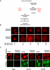
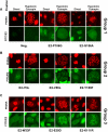
The papillomavirus E2 protein is required for viral transcriptional regulation, DNA replication and genome segregation. We have previously shown that the E2 transactivator protein and BPV1 genomes are associated with mitotic chromosomes; E2 links the genomes to cellular chromosomes to ensure efficient segregation to daughter nuclei. The transactivation domain of the E2 protein is necessary and sufficient for association of the E2 protein with mitotic chromosomes. To determine which residues of this 200-amino-acid domain are important for chromosomal interaction, E2 proteins with amino acid substitutions in each conserved residue of the transactivation domain were tested for their ability to associate with mitotic chromosomes. Chromatin binding was assessed by using immunofluorescence on both spread and directly fixed mitotic chromosomes. E2 proteins defective in the transactivation and replication functions were unable to associate with chromosomes, and those that were competent in these functions were attached to mitotic chromosomes. However, several mutated proteins that were defective for chromosomal interaction could associate with chromosomes after treatment with agents that promote protein folding or when cells were incubated at lower temperatures. These results indicate that precise folding of the E2 transactivation domain is crucial for its interaction with mitotic chromosomes and that this association can be modulated.
Figures


Similar articles
-
The mitotic chromosome binding activity of the papillomavirus E2 protein correlates with interaction with the cellular chromosomal protein, Brd4.J Virol. 2005 Apr;79(8):4806-18. doi: 10.1128/JVI.79.8.4806-4818.2005. J Virol. 2005. PMID: 15795266 Free PMC article.
-
Interaction of the papillomavirus E2 protein with mitotic chromosomes.Virology. 2000 Apr 25;270(1):124-34. doi: 10.1006/viro.2000.0265. Virology. 2000. PMID: 10772985
-
Analysis of chromatin attachment and partitioning functions of bovine papillomavirus type 1 E2 protein.J Virol. 2004 Feb;78(4):2100-13. doi: 10.1128/jvi.78.4.2100-2113.2004. J Virol. 2004. PMID: 14747575 Free PMC article.
-
Bovine papillomavirus type 1 genomes and the E2 transactivator protein are closely associated with mitotic chromatin.J Virol. 1998 Mar;72(3):2079-88. doi: 10.1128/JVI.72.3.2079-2088.1998. J Virol. 1998. PMID: 9499063 Free PMC article.
-
Brd4: tethering, segregation and beyond.Trends Microbiol. 2004 Dec;12(12):527-9. doi: 10.1016/j.tim.2004.10.002. Trends Microbiol. 2004. PMID: 15539109 Review.
Cited by
-
Replication-dependent and independent mechanisms for the chromosome-coupled persistence of a selfish genome.Nucleic Acids Res. 2016 Sep 30;44(17):8302-23. doi: 10.1093/nar/gkw694. Epub 2016 Aug 4. Nucleic Acids Res. 2016. PMID: 27492289 Free PMC article.
-
Variations in the association of papillomavirus E2 proteins with mitotic chromosomes.Proc Natl Acad Sci U S A. 2006 Jan 24;103(4):1047-52. doi: 10.1073/pnas.0507624103. Epub 2006 Jan 13. Proc Natl Acad Sci U S A. 2006. PMID: 16415162 Free PMC article.
-
Inhibition of E2 binding to Brd4 enhances viral genome loss and phenotypic reversion of bovine papillomavirus-transformed cells.J Virol. 2005 Dec;79(23):14956-61. doi: 10.1128/JVI.79.23.14956-14961.2005. J Virol. 2005. PMID: 16282494 Free PMC article.
-
Reconstitution of papillomavirus E2-mediated plasmid maintenance in Saccharomyces cerevisiae by the Brd4 bromodomain protein.Proc Natl Acad Sci U S A. 2005 Feb 22;102(8):2998-3003. doi: 10.1073/pnas.0407818102. Epub 2005 Feb 14. Proc Natl Acad Sci U S A. 2005. PMID: 15710895 Free PMC article.
-
The mitotic chromosome binding activity of the papillomavirus E2 protein correlates with interaction with the cellular chromosomal protein, Brd4.J Virol. 2005 Apr;79(8):4806-18. doi: 10.1128/JVI.79.8.4806-4818.2005. J Virol. 2005. PMID: 15795266 Free PMC article.
References
-
- Antson, A. A., J. E. Burns, O. V. Moroz, D. J. Scott, C. M. Sanders, I. B. Bronstein, G. G. Dodson, K. S. Wilson, and N. J. Maitland. 2000. Structure of the intact transactivation domain of the human papillomavirus E2 protein. Nature 403:805-809. - PubMed
-
- Baskakov, I., and D. W. Bolen. 1998. Forcing thermodynamically unfolded proteins to fold. J. Biol. Chem. 273:4831-4834. - PubMed
MeSH terms
Substances
LinkOut - more resources
Full Text Sources
Other Literature Sources

