

Blockade of the poliovirus-induced cytopathic effect in neural cells by monoclonal antibody against poliovirus or the human poliovirus receptor
- PMID: 15650178
- PMCID: PMC544096
- DOI: 10.1128/JVI.79.3.1523-1532.2005
Blockade of the poliovirus-induced cytopathic effect in neural cells by monoclonal antibody against poliovirus or the human poliovirus receptor
Abstract
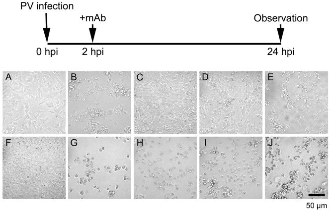
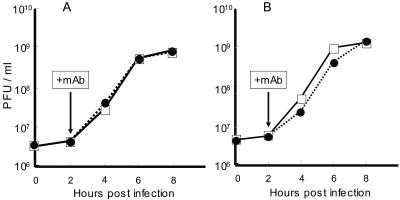
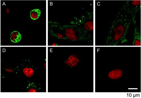
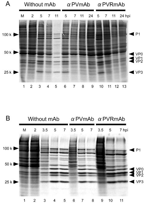
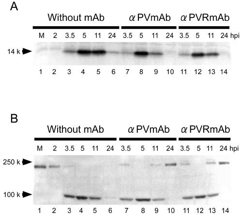
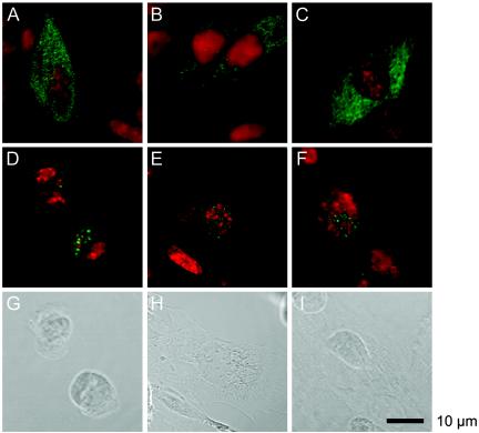
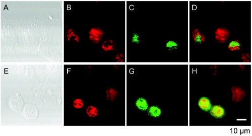
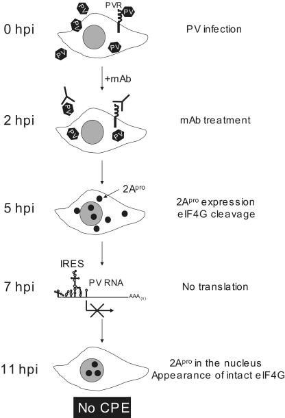
The poliovirus (PV)-induced cytopathic effect (CPE) was blocked in neural cells but not in HeLa cells by the addition of monoclonal antibody (MAb) against PV or the human PV receptor (CD155) 2 h postinfection (hpi). Since each MAb has the ability to block viral infection, no CPE in PV-infected neural cells appeared to result from the blockade of multiple rounds of viral replication. Pulse-labeling experiments revealed that virus-specific protein synthesis proceeded 5 hpi with or without MAbs. However, in contrast to the results obtained without MAbs, virus-specific protein synthesis with MAbs was not detected 7 hpi. Shutoff of host translation was also not observed in the presence of MAbs. Western blot analysis showed that 2Apro, the viral protein which mediates the cleavage of eukaryotic translation initiation factor eIF4G, was still present 11 hpi. However, intact eIF4G appeared 11 hpi. An immunocytochemical study indicated that 2Apro was detected only in the nucleus 11 hpi. These results suggest that neural cells possess protective response mechanisms against PV infection as follows: (i) upon PV infection, neural cells produce a factor(s) to suppress PV internal ribosome entry site activity by 7 hpi, (ii) a factor which supports cap-dependent translation for eIF4G may exist in infected cells when no intact eIF4G is detected, and (iii) the remaining 2Apro is not effective in cleaving eIF4G because it is imported into the nucleus by 11 hpi.
Figures
Similar articles
-
Studies on dicistronic polioviruses implicate viral proteinase 2Apro in RNA replication.Virology. 1993 Oct;196(2):739-47. doi: 10.1006/viro.1993.1531. Virology. 1993. PMID: 8396807
-
Persistent infection of human erythroblastoid cells by poliovirus.Virology. 1993 May;194(1):200-9. doi: 10.1006/viro.1993.1250. Virology. 1993. PMID: 8386873
-
Internal ribosomal entry site scanning of the poliovirus polyprotein: implications for proteolytic processing.Virology. 1998 Oct 10;250(1):241-53. doi: 10.1006/viro.1998.9376. Virology. 1998. PMID: 9770438
-
[Anti-poliovirus response of neural cells].Uirusu. 2003 Jun;53(1):41-9. doi: 10.2222/jsv.53.41. Uirusu. 2003. PMID: 14556501 Review. Japanese. No abstract available.
-
Molecular biology and cell-free synthesis of poliovirus.Biologicals. 1993 Dec;21(4):349-56. doi: 10.1006/biol.1993.1095. Biologicals. 1993. PMID: 8024750 Review.
Cited by
-
Scavenger receptor B2 is a cellular receptor for enterovirus 71.Nat Med. 2009 Jul;15(7):798-801. doi: 10.1038/nm.1992. Epub 2009 Jun 21. Nat Med. 2009. PMID: 19543282
-
Differential In Vitro Infection of Neural Cells by Astroviruses.mBio. 2019 Jul 9;10(4):e01455-19. doi: 10.1128/mBio.01455-19. mBio. 2019. PMID: 31289185 Free PMC article.
-
2A protease is not a prerequisite for poliovirus replication.J Virol. 2010 Jun;84(12):5947-57. doi: 10.1128/JVI.02575-09. Epub 2010 Apr 14. J Virol. 2010. PMID: 20392856 Free PMC article.
-
A second look at cellular mRNA sequences said to function as internal ribosome entry sites.Nucleic Acids Res. 2005 Nov 28;33(20):6593-602. doi: 10.1093/nar/gki958. Print 2005. Nucleic Acids Res. 2005. PMID: 16314320 Free PMC article. Review.
-
Characterization of the New World monkey homologues of human poliovirus receptor CD155.J Virol. 2008 Jul;82(14):7167-79. doi: 10.1128/JVI.02664-07. Epub 2008 May 14. J Virol. 2008. PMID: 18480448 Free PMC article.
References
-
- Aldabe, R., A. Barco, and L. Carrasco. 1996. Membrane permeabilization by poliovirus proteins 2B and 2BC. J. Biol. Chem. 271:23134-23137. - PubMed
-
- Bienz, K., D. Egger., Y. Rasser., and W. Bossart. 1980. Kinetics and location of poliovirus macromolecular synthesis in correlation to virus-induced cytopathology. Virology 100:390-399. - PubMed
Publication types
MeSH terms
Substances
LinkOut - more resources
Full Text Sources
Research Materials
Miscellaneous
