. 2005 Feb;79(3):1569-80.
doi: 10.1128/JVI.79.3.1569-1580.2005.

Hepatitis C virus (HCV) constitutively activates STAT-3 via oxidative stress: role of STAT-3 in HCV replication

Affiliations

Hepatitis C virus (HCV) constitutively activates STAT-3 via oxidative stress: role of STAT-3 in HCV replication

Gulam Waris et al. J Virol. 2005 Feb.

Retraction in

Abstract

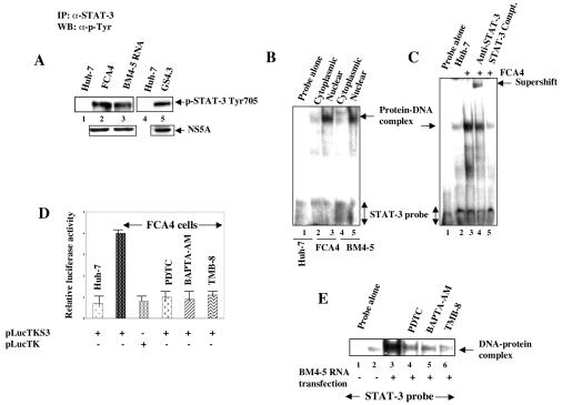
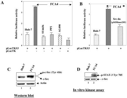
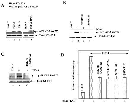
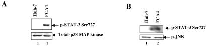
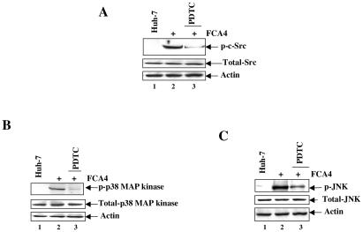
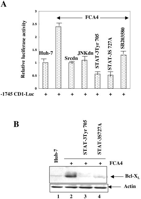
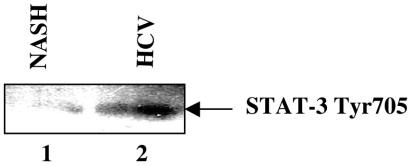
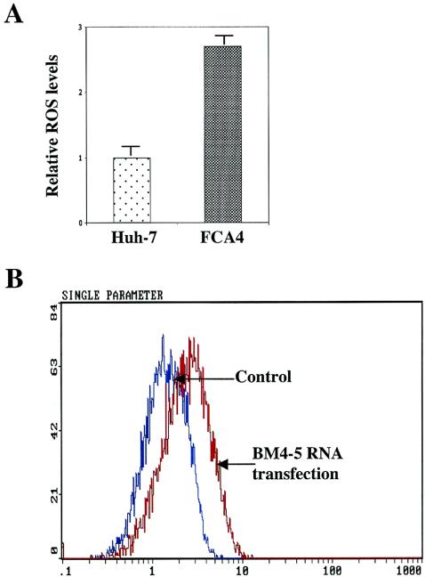
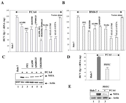
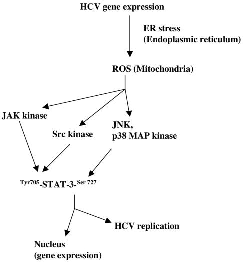
The hepatitis C virus (HCV) causes chronic hepatitis, which often results in liver cirrhosis and hepatocellular carcinoma. We have previously shown that HCV nonstructural proteins induce activation of STAT-3 via oxidative stress and Ca2+ signaling (G. Gong, G. Waris, R. Tanveer, and A. Siddiqui, Proc. Natl. Acad. Sci. USA 98:9599-9604, 2001). In this study, we focus on the signaling pathway leading to STAT-3 activation in response to oxidative stress induced by HCV translation and replication activities. Here, we demonstrate the constitutive activation of STAT-3 in HCV replicon-expressing cells. The HCV-induced STAT-3 activation was inhibited in the presence of antioxidant (pyrrolidine dithiocarbamate) and Ca2+ chelators (BAPTA-AM and TMB-8). Previous studies have shown that maximum STAT-3 transactivation requires Ser727 phosphorylation in addition to tyrosine phosphorylation. Using a series of inhibitors and dominant negative mutants, we show that HCV-induced activation of STAT-3 is mediated by oxidative stress and influenced by the activation of cellular kinases, including p38 mitogen-activated protein kinase, JNK, JAK-2, and Src. Our results also suggest a potential role of STAT-3 in HCV RNA replication. We also observed the constitutive activation of STAT-3 in the liver biopsy of an HCV-infected patient. These studies provide an insight into the mechanisms by which HCV induces intracellular events relevant to liver pathogenesis associated with the viral infection.


Figures

FIG. 1.
FIG. 1.
HCV replicons induce tyrosine phosphorylation of STAT-3. (A) Whole-cell lysates from cells expressing HCV replicons were immunoprecipitated with anti-STAT-3 serum, fractionated by SDS-PAGE, and immunoblotted with antiphosphotyrosine monoclonal antibody. Lanes 1 and 4, untransfected Huh-7 lysates; lanes 2 and 5, FCA4 and GS4.3 cells expressing HCV subgenomic replicons; lane 3, Huh-7 cells transfected with in vitro-synthesized BM4-5 RNA. The bottom panel represents the expression of HCV NS5A in HCV replicon-expressing cells. (B) An EMSA was carried out in the presence of γ-32P-labeled STAT-3 cognate nucleotide probe, and the nuclear lysates were prepared from FCA4 cells and Huh-7 cells transiently transfected with in vitro-synthesized BM4-5 RNA. Lane 1, STAT-3 probe incubated with Huh-7 nuclear lysates; lanes 2 and 3, equal amounts of FCA4 cytoplasmic and nuclear lysates, respectively; lanes 4 and 5, equal amounts of BM4-5 RNA-transfected cytoplasmic and nuclear lysates, respectively. (C) EMSA. Lane 1, probe alone; lanes 2 and 3, equal amounts of Huh-7 and FCA4 nuclear lysates; lane 4, DNA-protein complex incubated with anti-STAT-3 serum; lane 5, DNA-protein complex treated with a 100-fold excess of unlabeled STAT-3 oligonucleotide. (D) Luciferase reporter gene assay. FCA4 cells were transfected with STAT-3-responsive pLucTKS3 and pLucTK (without STAT-3 binding sites) luciferase plasmids. At 36 h posttransfection, cells were treated with PDTC (6 h), BAPTA-AM (2 h), and TMB-8 (4 h) at various times before preparing the lysates for luciferase activity. (E) EMSA. Lane 1, probe alone; lanes 2 and 3, equal amounts of untransfected and BM4-5 replicon RNA-transfected nuclear lysates; lanes 4, 5, and 6, BM4-5 RNA-transfected lysates treated with PDTC (6 h), BAPTA-AM (2 h), and TMB-8 (4 h) at various times.
FIG. 2.
FIG. 2.
HCV replicon-induced activation of STAT-3 is mediated by JAK2 and Src kinases. (A) Huh-7 and FCA4 cells were transfected with STAT-3-responsive pLucTKS3 luciferase plasmid. At 36 h posttransfection, cells were treated with inhibitors of Src kinase (10 μM SU6656 for 2 h and 20 μM PP2 for 2 h) and JAK2 (40 μM AG490 for 4 h) before preparing the lysates for luciferase activity determinations. (B) FCA4 cells were transfected with a Src dominant negative (pM5Hmet295) expression vector along with the STAT-3-responsive pLucTKS3 luciferase plasmid. At 36 h posttransfection, cellular lysates were prepared for luciferase activity determinations. (C) HCV replicon activates Src kinase. Equal amounts of cellular lysates from Huh-7 and FCA4 cells were subjected to SDS-PAGE and immunoblotted with anti-Src serum. Lane 1, Huh-7 lysates; lane 2, FCA4 lysates. (D) In vitro c-Src kinase assay. Equal amounts of cellular lysates were immunoprecipitated with anti-Src serum, and Src activity was measured in an in vitro kinase assay using [γ-32P]ATP and unphosphorylated STAT-3 as a substrate. Labeled STAT-3 was resolved by SDS-PAGE and visualized by autoradiography. Lane 1, Huh-7 lysates; lane 2, FCA4 lysates. The bottom panel represents the total Src activity in Huh-7 and FCA4 lysates.
FIG. 3.
FIG. 3.
HCV replicon stimulates phosphorylation of STAT-3 at Ser727. (A) Whole-cell lysates from cells expressing HCV replicons were immunoprecipitated with anti-STAT-3 serum, fractionated by SDS-PAGE, and immunoblotted with anti-STAT-3 Ser727 serum. Lanes 1 and 4, untransfected lysates; lanes 2 and 3, FCA4 and GS4.3 cells expressing HCV subgenomic replicons; lane 5, transfected with in vitro-synthesized BM4-5 RNA. The bottom panel represents the total STAT-3 in Huh-7 and HCV replicon-expressing cells. (B) FCA4 cells were treated with inhibitors of p38 MAPK (10 μM SB203580 for 6 h) and JNK (30 μM SP600125 for 2 h). Equal amounts of cellular lysates were immunoprecipitated with anti-STAT-3 serum and immunoblotted with anti-STAT-3 Ser727 serum. Lanes 1 and 2, equal amounts of Huh-7 and FCA4 lysates; lanes 3 and 4, FCA4 lysates treated with SB203580 and SP600125, respectively. (C) FCA4 cells were transfected with a JNK dominant negative (p54JNK2α) expression vector. Equal amounts of cellular lysates were immunoprecipitated with anti-STAT-3 serum and immunoblotted with anti-STAT-3 Ser727 serum. Lanes 1 and 2, equal amounts of Huh-7 and FCA4 lysates; lane 3, FCA4 lysates expressing dominant negative JNK. (D) Huh-7 and FCA4 cells were transfected with the STAT-3-responsive pLucTKS3 luciferase plasmid along with dominant negative mutants of JNK (p54JNK2α) and STAT-3 (STAT-3S727A). At 36 h posttransfection, FCA4 cells expressing pLucTKS3 alone were treated with inhibitors of p38 MAPK (SB203580) and JNK (SP600125) at various times before preparing the lysates for luciferase activity assays.
FIG. 4.
FIG. 4.
HCV replicon activates MAPKs. Equal amounts of cellular lysates were immunoprecipitated with anti-p38 MAPK and anti-phospho-JNK antibodies, and the kinase activities were measured in an in vitro kinase assay using [γ-32P]ATP and unphosphorylated STAT-3 as substrate. Labeled STAT-3 was resolved by SDS-PAGE and visualized by autoradiography. Lanes 1, Huh-7 lysates; lanes 2, FCA4 lysates. The bottom panel represents the total p38 MAPK and phospho-JNK activities in Huh-7 and FCA4 lysates, respectively.
FIG. 5.
FIG. 5.
HCV replicon-induced c-Src, p38 MAPK, and JNK are sensitive to antioxidants. Huh-7 and FCA4 cells were treated with the antioxidant PDTC (100 μM) for 6 h. Equal amounts of cellular lysates were subjected to SDS-PAGE and immunoblotted with anti-phospho-Src, anti-phospho-p38 MAPK, and anti-phospho-JNK antibodies. Lanes 1, Huh-7 lysates; lanes 2, FCA4 lysates; lanes 3, FCA4 lysates treated with antioxidant (PDTC) for 6 h before harvesting the cells.
FIG. 6.
FIG. 6.
HCV replicon induces cyclin D1 and Bcl-XL expression. (A) Huh-7 and FCA4 cells were transiently transfected with cyclin D1 luciferase reporter along with the dominant negative mutants of Src (pM5Hmet295), JNK (p54JNK2α), and STAT-3 (pGS5hSTAT-3β; STAT-3S727A). At 36 h posttransfection, cellular lysates were prepared for luciferase activity assays. (B) Equal amounts of Huh-7 and FCA4 lysates expressing dominant negative mutants of STAT-3 were subjected to SDS-PAGE and immunoblotted with anti-Bcl-XL serum. Lane 1, Huh-7 lysates; lane 2, FCA4 lysates; lanes 3 and 4, FCA4 lysates expressing STAT-3 dominant negative mutants.
FIG. 7.
FIG. 7.
Constitutive activation of STAT-3 in the liver of HCV-positive patients. Cellular extracts were prepared from liver biopsies of HCV-infected and uninfected (NASH) patients as described in Materials and Methods. Equal amounts of cellular extracts were subjected to SDS-PAGE and electroblotted onto a nitrocellulose membrane. The membrane was then immunoblotted with anti-phospho-STAT-3 antibody. Lane 1, cellular lysates from NASH; lane 2, cellular lysates from HCV-infected patients.
FIG. 8.
FIG. 8.
HCV replicon induces intracellular production of ROS. (A) Huh-7 and FCA4 cells were treated with 4 μM DHE for 45 min. Cells were harvested, and ROS levels were measured by flow cytometry with excitation emission at 605 nm. The bars show the fold increase in oxidized DHE fluorescence. (B) Untransfected (control) or in vitro-synthesized BM4-5 RNA-transfected Huh-7 cells were treated with 4 μM DHE. ROS levels were assessed in untransfected cells (blue) and cells transfected with BM4-5 RNA (red).
FIG. 9.
FIG. 9.
Effect of STAT-3 on HCV RNA replication. (A, B, and D) FCA4 cells and Huh-7 cells transiently transfected with in vitro-synthesized BM4-5 RNA were first incubated with the inhibitors of tyrosine and MAP kinases (AG490, PP2, SB203580, and SP600125) and the antioxidant PDTC overnight. The replicon-expressing cells were also transiently transfected with the dominant negative expression vectors of Src (pM5Hmet295), STAT-3 Tyr705 (pSG5hSTAT-3β), and STAT-3 S727A. The total RNA was extracted and subjected to quantitative RT-PCR analysis. The data are expressed as relative HCV RNA levels in relation to the RNA levels in the control cells. (C and E) FCA4 cells were treated with the inhibitors of tyrosine and MAP kinases (AG490, PP2, SB203580, and SP600125) and the antioxidant PDTC overnight. Equal amounts of cellular lysates were subjected to SDS-PAGE and Western blotted with anti-NS5A serum. (C) Lanes 1 and 2, equal amounts of Huh-7 and FCA4 lysates; lane 3, 4, 5, and 6, FCA4 lysates treated with PP2, AG490, SP600125, and SB203580, respectively. (E) Lanes 1 and 2, equal amounts of Huh-7 and FCA4 lysates; lane 3, FCA4 lysates treated with PDTC.
FIG. 10.
FIG. 10.
Model illustrating the mechanism(s) of HCV replicon-induced activation of STAT-3 via oxidative stress. This pathway involves the activation of tyrosine and MAP kinases in HCV replicon-expressing cells. By a mechanism not clearly understood, STAT-3 enhances HCV RNA replication.

Similar articles

Cited by

References

    1. Abe, J., M. Takahashi, M. Ishida, J. W. Lee, and B. C. Berk. 1997. c-Src is required for oxidative stress-mediated activation of big mitogen-activated protein kinase 1 (BMK1). J. Biol. Chem. 272:20389-20394. - PubMed
    1. Bartenschlager, R., and V. Lohmann. 2000. Replication of hepatitis C virus. J. Gen. Virol. 81:1631-1648. - PubMed
    1. Bartenschlager, R., and V. Lohmann. 2001. Novel cell culture systems for the hepatitis C virus. Antivir. Res. 52:1-17. - PubMed
    1. Bowman, T., R. Garcia, J. Turkson, and R. Jove. 2000. STATs in oncogenesis. Oncogene 19:2474-2488. - PubMed
    1. Bowman, T., M. A. Broome, D. Sinibaldi, W. Wharton, W. J. Pledger, J. M. Sedivy, R. Irby, T. Yeatman, S. A. Courtneidge, and R. Jove. 2001. STAT-3 mediated myc expression is required for Src transformation and PDGF-induced mitogenesis. Proc. Natl. Acad. Sci. USA 98:7319-7324. - PMC - PubMed

Publication types

MeSH terms