. 2005 Feb;79(3):1613-22.
doi: 10.1128/JVI.79.3.1613-1622.2005.

Efficient inhibition of hepatitis B virus infection by acylated peptides derived from the large viral surface protein

Affiliations

Efficient inhibition of hepatitis B virus infection by acylated peptides derived from the large viral surface protein

Philippe Gripon et al. J Virol. 2005 Feb.

Abstract

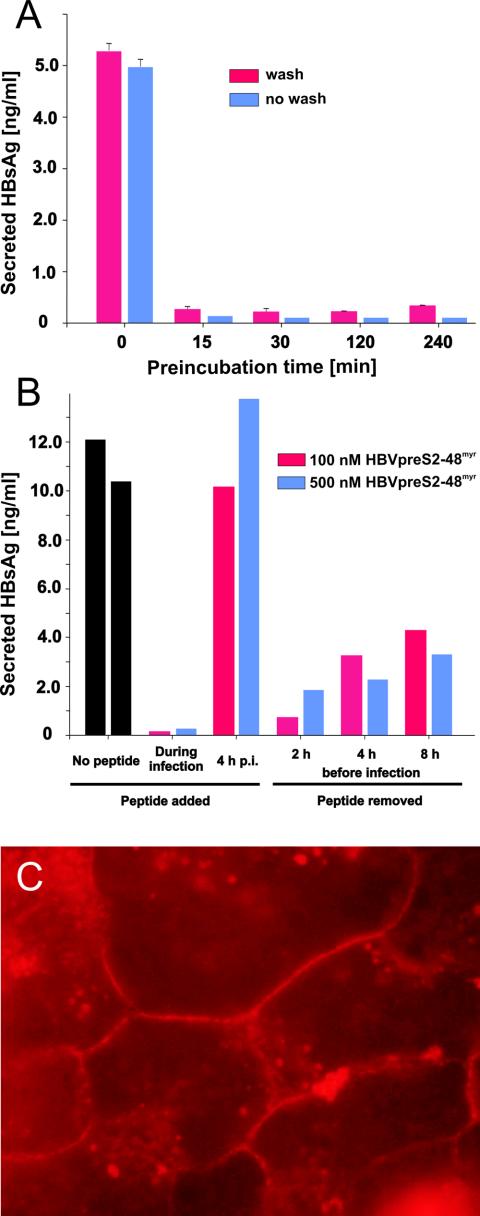
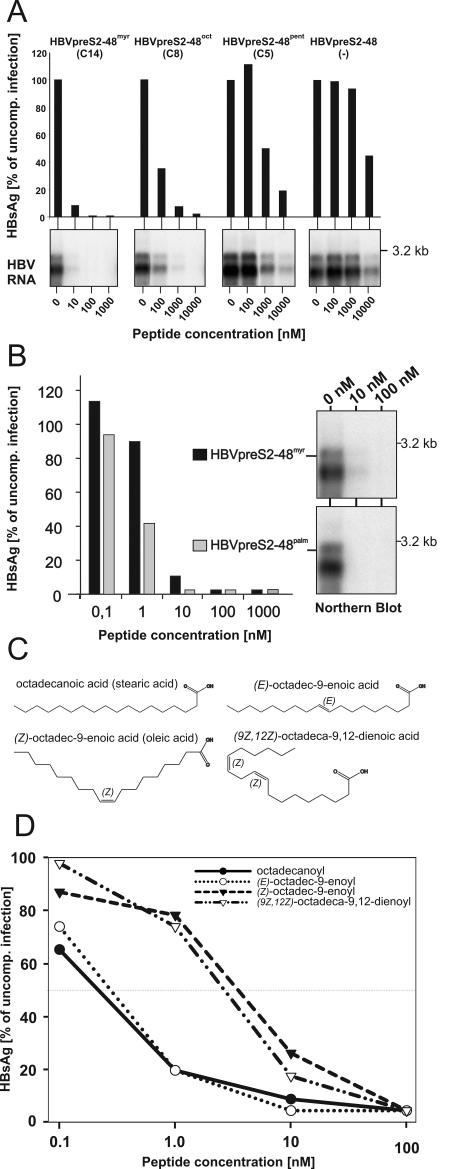
The lack of an appropriate in vitro infection system for the major human pathogen hepatitis B virus (HBV) has prevented a molecular understanding of the early infection events of HBV. We used the novel HBV-infectible cell line HepaRG and primary human hepatocytes to investigate the interference of infection by HBV envelope protein-derived peptides. We found that a peptide consisting of the authentically myristoylated N-terminal 47 amino acids of the pre-S1 domain of the large viral envelope protein (L protein) specifically prevented HBV infection, with a 50% inhibitory concentration (IC50) of 8 nM. The replacement of myristic acid with other hydrophobic moieties resulted in changes in the inhibitory activity, most notably by a decrease in the IC50 to picomolar concentrations for longer unbranched fatty acids. The obstruction of HepaRG cell susceptibility to HBV infection after short preincubation times with the peptides suggested that the peptides efficiently target and inactivate a receptor at the hepatocyte surface. Our data both shed light on the molecular mechanism of HBV entry into hepatocytes and provide a basis for the development of potent hepadnaviral entry inhibitors as a novel therapeutic concept for the treatment of hepatitis Beta.


Figures

FIG. 1.
FIG. 1.
Infection inhibition activity of terminally deleted myristoylated HBV pre-S1 peptides. (A) Schematic illustration of HBV L-protein-derived pre-S1 peptides used for infection inhibition. HBVpreS/2-78myr, consisting of the first 77 amino acids of the HBV pre-S1 domain (subtype ayw) as an N-terminal amide of myristic acid (11), and progressively C-terminally deleted myristoylated variants aredepicted on an equivalent scale. Numbers in the names of the peptides specify the first and last amino acids; “myr” indicates that the respective peptide is N-terminally myristoylated at glycine 2. (B) Competition of HBV infection by two myristoylated pre-S1 peptides, HBVpreS/2-68myr and HBVpreS/2-48myr. HepaRG cells were infected with a multiplicity of genome equivalents of ≈50 in the presence of 0.8, 8, 80, and 800 nM concentrations of the respective pre-S peptides. Fourteen hours later, the cells were washed twice with culture medium. At 12 days postinfection, cellular mRNAs were prepared and analyzed by Northern blotting as described previously (11). Both genomic and subgenomic RNAs are represented. The specificity of infection was controlled by an RNA analysis of uninfected HepaRG cells (left lane, no virus); the specificity of inhibition was tested by the addition of the myristoylated DHBV pre-S peptide DHBVpreS/2-41myr at 800 nM (control). RNA size markers are shown on the right. (C) PHH and HepaRG cells were infected overnight under comparable conditions in the presence (left) or absence (right) of PEG, with 0, 10, or 100 nM HBVpreS2/48myr as an inhibitor. After removal of the peptide, the medium was exchanged every 2 to 3 days and the amount of HBsAg was determined from days 12 to 14 postinfection. Error bars in the uncompeted infections represent the standard deviations for six independent infections. (D) HepaRG cells were infected in the presence of 0.8 μM (left) and 8 μM (right) HBVpreS/2-8myr, HBVpreS/2-18myr, HBVpreS/2-28myr, HBVpreS/2-39myr, and as controls, HBVpreS/2-48myr and DHBVpreS/2-41myr. The infectious inoculum and the peptides were incubated overnight, washed, and replaced with new medium. At 12 days postinfection, the collected cell culture supernatants from days 8 to 12 were analyzed for secreted HBsAg by use of a quantitative commercially available ELISA. The results obtained are given in nanograms of HBsAg per milliliter of cell culture medium. (E) Comparative HBV infection assay with the two myristoylated pre-S1-derived peptides HBVpreS19-48myr, which lacks the first 17 pre-S1 amino acids and carries an artificial myristic acid residue at the N-terminal leucine 19, and HBVpreS/2-48myr. HepaRG cells were infected with HBV (MGE = 50) for 12 h in the presence of 8, 80, and 800 nM concentrations of the respective peptide. The medium was changed, and newly synthesized HBsAg was determined between days 8 and 12 postinfection. Values are given as relative units (optical densities) obtained from the ELISA reader.
FIG. 2.
FIG. 2.
HBV infection inhibition by a pre-S-derived peptide of the WMHBV L protein. (A) Amino acid sequence alignment of the two pre-S1-derived peptides HBVpreS/2-48myr (H) and WMHBVpreS/2-48myr (W). Numbers above the sequences indicate amino acid positions; gray boxes denote perfect sequence accordance. The sequence identity within this pre-S sequence was 64%. Note that most of the remaining 36% amino acid deviations are nonconservative. (B) Comparative HBV infection competition assay with 0.8, 8, 80, and 800 nM WMHBVpreS/2-48myr (left) and HBVpreS/2-48myr (right). At 12 days postinoculation of virus and peptide, total cellular mRNAs were isolated and viral transcripts (genomic and subgenomic RNAs) were analyzed by Northern blot hybridization using an HBV-specific probe. Size markers are indicated on the right. (C) HBV infection competition using nonmyristoylated pre-S-derived peptides of DHBV (DHBVpreS/1-41), WMHBV (WMHBVpreS/1-48), and HBV (HBVpreS/1-48) at the elevated concentrations of 25 μM (middle) and 100 μM (right) in comparison to an uncompeted infection (left lane in left panel) and a mock infection (right lane in left panel). Cells were incubated with HBV in the presence of the indicated peptide concentrations for 16 h. Twelve days after the removal of the inoculum, the cells were washed twice, RNAs were prepared, and viral transcripts were detected by Northern blot hybridization.
FIG. 3.
FIG. 3.
Epitopes of neutralizing anti-pre-S1 monoclonal antibodies overlap with key amino acids required for efficient infection inhibition. (A) Schematic drawing of pre-S1 and pre-S2 domains of the HBV L protein (subtype ayw). Numbers at the top indicate the N-terminally myristoylated glycine 2 of pre-S1, the last amino acid of pre-S1 (108), and pre-S2 (163). The position of the inhibitory domain and the sequence of the wild-type inhibitory peptide HBVpreS/2-48myr are depicted below, with the recognition epitopes for the two neutralizing monoclonal antibodies MA18/7 (20-DPAF-23) and 5a19 (26-NTANPDW-32) highlighted in bold. The sequences of the deletion mutants HBVpreS/2-48myrΔ20-21, HBVpreS/2-48myrΔ20-23, and HBVpreS/2-48myrΔ23-27 are aligned with the HBVpreS/2-48myr peptide sequence. (B) Comparative HBV infection competition assay using the internal deletion mutants depicted in panel A. HepaRG cells were infected either in the absence (0 nM) or in the presence of 10, 100, and 1,000 nM HBVpreS/2-48myr, HHBVpreS/2-44myr (a heron hepatitis B virus-derived analogue that served as an additional control [32]), HBVpreS/2-48myrΔ20-21, HBVpreS/2-48myrΔ20-23, and HBVpreS/2-48myrΔ23-27. The infectious inoculum and the peptides were incubated overnight. After being washed, the cells were maintained for another 12 days to allow viral gene expression. Cell culture supernatants from days 8 to 12 were collected and analyzed for secreted HBsAg by use of a quantitative commercially available ELISA. HBsAg values from the respective uncompeted infection were set to 100%, and the degree of infection inhibition is given as a percentage of the uncompeted infection. The absolute mean value of HBsAg for the uncompeted infection was 6.3 ng/ml.
FIG. 4.
FIG. 4.
HBVpreS/2-48myr addresses a cell-associated factor on the surfaces of hepatocytes. (A) HBV infection inhibition after preincubation of HepaRG cells with HBVpreS/2-48myr. HepaRG cells were incubated with HBVpreS/2-48myr (800 nM) for 0, 15, 30, 120, and 240 min. Subsequently, the peptide was either removed (red bars) or lefton the cells (blue bars) for the duration of the HBV infection (an additional 12 h with ≈50 genome equivalents per cell). After being washed, the cells were incubated for an additional 12 days to allow the expression of viral genes. For quantification, cell culture supernatants were collected between days 8 and 12 postinfection and then subjected to a quantitative HBsAg ELISA. Experiments were done in duplicate; standard deviations are indicated by error bars. (B) Effect of HBVpreS/2-48myr on HBV infection when administered before or after the establishment of infection. HepaRG cells were inoculated overnight with ≈50 genome equivalents of HBV in the absence or presence of 100 nM (red bars) or 500 nM (blue bars) HBVpreS/2-48myr. In contrast, HBVpreS/2-48myr was added just 4 h after removal of the infectious inoculum and was then left on the cells for an additional 16 h. Alternatively, HBVpreS/2-48myr was incubated with HepaRG cells at the indicated concentrations (incubation time, 16 h). After removal of the peptide, the cells were cultivated for another 2, 4, or 8 h at 37°C. Subsequently, the cells were infected with HBV in the absence of any inhibitor (12 h; MGE, ≈50) and then washed, and virus replication was allowed to occur for an additional 12 days. As described above, newly synthesized HBsAg was determined by quantitative HBsAg ELISA. (C) Direct visualization of HBVpreS/2-48myr binding to PHH. HBVpreS/2-48myr was labeled with Cy3 as described in Materials and Methods. The labeled peptide was allowed to bind PHH for 1 h. After removal of the peptide, the cells were fixed and analyzed by fluorescence microscopy.
FIG. 5.
FIG. 5.
Influence of the nature of the N-terminal acyl moiety on pre-S-specific HBV infection inhibition. (A) HBV infection inhibition assay using HBVpreS/2-48 variants without or with shortened hydrocarbon acid chain lengths, i.e., HBVpreS/2-48, pentanoyl-HBVpreS/2-48 (HBVpreS/2-48pent [C5]), octanoyl-HBVpreS/2-48 (HBVpreS/2-48oct [C8]), and as a reference, myristoyl-HBVpreS/2-48 (HBVpreS/2-48myr [C14]). HepaRG cells were infected overnight in the presence of the indicated concentrations of the respective acyl HBV pre-S inhibitor. After removal of the inoculum, the cells were further cultivated and the expression of newly synthesized HBsAg between days 8and 12 (bar diagrams) and of viral transcripts on day 12 (autoradiograms) was analyzed by ELISA and Northern blot hybridization. Note the 10-fold higher concentration used for the inhibition of shortened variants. (B) Comparative HBV infection inhibition assay using HBVpreS/2-48myr and its derivative palmitoyl-HBVpreS/2-48 (HBVpreS/2-48palm [C16]) with a lengthened hydrocarbon acid chain. HepaRG cells were infected in parallel for 12 h in the presence of 0.1, 1, 10, 100, and 1,000 nM concentrations of the two peptides. After being washed, the cells were cultivated for another 12 days and the expression of HBsAg between days 8 and 12 was quantified by ELISA (bar diagram). Values are displayed as percentages of the corresponding uncompeted control infection. In addition to HBsAg secretion, viral RNAs prepared at 12 days postinfection were subjected to Northern blot analysis (autoradiograms). Data for genomic and subgenomic RNAs are shown. (C) Structural formulas of octadecanoic (stearic) acid, (Z)-octadec-9-enoic (oleic) acid, (E)-octadec-9-enoic acid, and (9Z,12Z)-octadeca-9,12-dienoic acid, which were used for the synthesis of the corresponding HBVpreS/2-48 peptides analyzed below. Note the steric differences of the two Z derivatives compared to the saturated and singly unsaturated E variants. (D) Comparative HBV infection competition assay using the four acylated pre-S2-48 derivatives depicted in panel C. HepaRG cells were inoculated overnight with HBV (≈50 genome equivalents) in the presence of 0.1, 1, 10, and 100 nM concentrations of the respective peptide derivatives. Cells were washed, and newly synthesized HBsAg (days 8 to 12) was quantified by ELISA. Values are given as percentages of the corresponding uncompeted control infection.

Similar articles

Cited by

References

    1. Acs, G., M. A. Sells, R. H. Purcell, P. Price, R. Engle, M. Shapiro, and H. Popper. 1987. Hepatitis B virus produced by transfected HepG2 cells causes hepatitis in chimpanzees. Proc. Natl. Acad. Sci. USA 84:4641-4644. - PMC - PubMed
    1. Berting, A., C. Fischer, S. Schaefer, W. Garten, H. D. Klenk, and W. H. Gerlich. 2000. Hemifusion activity of a chimeric influenza virus hemagglutinin with a putative fusion peptide from hepatitis B virus. Virus Res. 68:35-49. - PubMed
    1. Bruss, V., and D. Ganem. 1991. The role of envelope proteins in hepatitis B virus assembly. Proc. Natl. Acad. Sci. USA 88:1059-1063. - PMC - PubMed
    1. Bruss, V., J. Hagelstein, E. Gerhardt, and P. R. Galle. 1996. Myristylation of the large surface protein is required for hepatitis B virus in vitro infectivity. Virology 218:396-399. - PubMed
    1. Chang, C. M., K. S. Jeng, C. P. Hu, S. J. Lo, T. S. Su, L. P. Ting, C. K. Chou, S. H. Han, E. Pfaff, J. Salfeld, et al. 1987. Production of hepatitis B virus in vitro by transient expression of cloned HBV DNA in a hepatoma cell line. EMBO J. 6:675-680. - PMC - PubMed

Publication types

MeSH terms