. 2005 Feb;79(3):1861-70.
doi: 10.1128/JVI.79.3.1861-1870.2005.

Glc1.8 from Microplitis demolitor bracovirus induces a loss of adhesion and phagocytosis in insect high five and S2 cells

Affiliations

Glc1.8 from Microplitis demolitor bracovirus induces a loss of adhesion and phagocytosis in insect high five and S2 cells

Markus Beck et al. J Virol. 2005 Feb.

Abstract

Polydnaviridae is a unique family of DNA viruses that are symbiotically associated with parasitoid wasps. Upon oviposition, wasps inject these viruses into their hosts, where they cause several physiological alterations, including suppression of the cellular immune response. Here we report that expression of the glc1.8 gene from Microplitis demolitor bracovirus (MdBV) causes a loss of adhesion by two hemocyte-like cell lines, namely, High Five cells from the lepidopteran Trichoplusia ni and S2 cells from the dipteran Drosophila melanogaster. The expression of recombinant Glc1.8 also greatly reduced the ability of these cells to phagocytize foreign targets. Glc1.8 is characterized by a signal peptide at its N terminus, an extracellular domain comprised of five nearly perfect tandem repeats of 78 amino acids, and a C-terminal hydrophobic domain that encodes a putative membrane anchor sequence. The expression of a Glc1.8 mutant lacking the anchor sequence resulted in a secreted protein that had no effect on adhesion or phagocytosis. In contrast, sequential deletion of the repeats in the extracellular domain resulted in a progressive reduction in immunosuppressive activity. Since each repeat and its associated glycosylation sites are nearly identical, these results suggested that adhesion-blocking activity depends more on the overall number of repeats in the extracellular domain than on the specific determinants within each repeat. While it severely compromised adhesion and phagocytic functions, Glc1.8 did not cause cell death. Collectively, these results indicate that Glc1.8 is a major pathogenic determinant of MdBV that is involved in suppression of the insect cellular immune response.


Figures

FIG. 1.
FIG. 1.
Structural domains of glc1.8 and deletion mutants produced for use in adhesion and phagocytosis assays. The top panel illustrates the major motifs and selected restriction enzyme sites in the previously published sequence of glc1.8 (GenBank accession no. AF267175). The signal peptide region is represented by a solid box, the five tandemly arrayed direct repeats are shown as gray boxes, and the transmembrane domain at the 3′ end is shown as a striped box. AflII, NsiI, SacII, and XbaI sites are marked by arrows. The bottom panel presents schematics for the constructs used in experiments. Glc1.8, wild-type protein. AflII digestion produced mutants (mutΔ1 to mutΔ4) with four, three, two, or one of the tandem repeats that comprise the extracellular domain of wild-type Glc1.8. A deletion mutant lacking the transmembrane domain (mutΔC) was produced by NsiI and SacII digestion of glc1.8.
FIG. 2.
FIG. 2.
Loss of adhesion by High Five cells is dose dependent and persistent. Phase-contrast micrographs show cells at 36 h posttransfection with pIZT/V5-His (empty vector) (A) and pIZT/Glc1.8 (B). Most cells in panel A are adhesive and have a fibroblastic morphology, whereas the cells in panel B are nonadhesive and rounded. Epifluorescence micrographs show cells at 48 h posttransfection with 2 μg of pIZT/Glc1.8 (C) or at 48 h postinfection with 0.1 wasp equivalents of MdBV (D). Glc1.8 (red) localized to the cell surface in both treatments, as visualized by the labeling of living cells with a mouse anti-Glc1.8 monoclonal antibody and a Texas Red-conjugated secondary antibody. Bar, 25 μm. (E) Effect of recombinant Glc1.8 expression on adhesion of High Five cells. Cells were transfected with 0.2 to 8 μg of pIZT/Glc1.8 per ml, and the numbers of cells expressing Glc1.8 that adhered to the surfaces of culture plates were counted 48 h later. Cells transfected with 2 μg of empty pIZT/V5-His vector per ml (vector) served as a negative control, while cells infected with 0.1 wasp equivalents of MdBV served as a positive control. The results are given as means ± standard errors (SE) for adhesive cells relative to the total number of cells (200) counted per sample (*, significantly different [P < 0.05] compared to the vector-only control by the SNK multiple comparison procedure; n = 6 replicates per treatment). (F) Persistence of nonadhesive phenotype and viability of High Five cells after transfection with 2 μg of pIZT/Glc1.8 per ml. The results are presented as means ± SE for adhesive cells relative to the total number of cells (200) counted per sample from 0 to 120 h posttransfection (n = 3 replicates per time point).
FIG. 3.
FIG. 3.
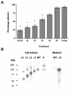
Loss of adhesion by High Five cells requires the transmembrane and glycosylated extracellular domains of Glc1.8. (A) The mutant mutΔC, which lacks the hydrophobic domain, had no effect on adhesion, whereas sequential deletion of the tandem repeats (Δ1 to Δ4) of the extracellular domain resulted in a progressive increase in the nonadhesive phenotype. Expression of the wild-type protein (Glc1.8) served as a positive control, while cells transfected with the empty pIZT/V5-His vector (vector) served as a negative control. Cells for all treatments were transfected with 2 μg of plasmid/ml and were assessed for the loss of adhesion 48 h later. The results are given as means ± SE for adhesive cells relative to the total number of cells (200) counted per sample (*, significantly different [P < 0.05] compared to the vector-only control by the SNK multiple comparison procedure; n = 6 replicates per treatment). (B) Lysates from cells and conditioned medium harvested at 48 h posttransfection were subjected to SDS-8 to 16% gradient PAGE, transferred to nitrocellulose, and immunoblotted, followed by visualization by chemiluminescence. The deletion mutants (Δ1 to Δ4 and ΔC) and wild-type Glc1.8 (WT) were detected with a mouse anti-V5 antibody specific for the C-terminal V5 epitope. Glc1.8 expressed from cells infected with 0.1 wasp equivalents of MdBV (V) was detected with an anti-Glc1.8 antibody.
FIG. 4.
FIG. 4.
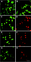
Disruption of actin cytoskeleton in High Five cells expressing Glc1.8. Normal uninfected cells (A) and cells transfected with the empty pIZT/V5-His vector (B) displayed elongated actin filaments (stress fibers) throughout the cells, as visualized by staining with Alexa 488-phalloidin. Cells infected by MdBV were visualized to show F-actin (C) or Glc1.8 (D) expression. These cells exhibit a rounded morphology and anaccumulation of F-actin around the periphery (green). Cells transfected with wild-type Glc1.8 were visualized to show actin (E) or Glc1.8 (F) expression. These cells also showed an accumulation of peripheral actin. Cells transfected with mutΔ4 were visualized to show actin (G) or Glc1.8 (H) expression. These cells showed intermediate modifications of the actin cytoskeleton even though Glc1.8 was detected on the cell surfaces (H). Cells for each treatment were infected with 0.1 wasp equivalents of MdBV or were transfected with 2 μg of plasmid/ml. Forty-eight hours later, the cells were fixed and permeabilized, and the actin cytoskeleton was visualized by the use of Alexa 488-phalloidin. Glc1.8 expression was visualized with anti-Glc1.8 and a Texas Red-conjugated secondary antibody. The cells were examined by confocal microscopy. Bar, 25 μm.
FIG. 5.
FIG. 5.
Glc1.8 expression induces loss of adhesion by S2 cells. (A) Cells were transfected with 2 μg of Glc1.8, the mutΔ1 mutant, the mutΔC mutant, or the empty pIZT/V5-His vector (vector)/ml and then assessed for the loss of adhesion 48 h later. The results are given as means ± SE for adhesive cells relative to the total number of cells (200) counted per sample (*, significantly different [P < 0.05] compared to the vector-only control by the SNK multiple comparison procedure; n = 8 replicates per treatment). Phase-contrast (B) and epifluorescence (C) micrographs of S2 cells were taken at 48 h posttransfection with wild-type Glc1.8. Most cells expressing Glc1.8 on their surfaces were rounded and nonadhesive. Living cells were labeled with an anti-Glc1.8 antibody and a Texas Red-conjugated secondary antibody. Bar, 50 μm.
FIG. 6.
FIG. 6.
Glc1.8 reduces phagocytosis of E. coli and polystyrene beads. (A and B) Influence of MdBV infection or Glc1.8 expression on adherence and phagocytosis by High Five cells. (C and D) Influence of Glc1.8 expression on adherence and phagocytosis by S2 cells. Cells were infected with 0.1 wasp equivalents of MdBV or transfected with wild-type Glc1.8, the mutΔ1 mutant, the mutΔC mutant, or the empty pIZT/V5-His vector (vector) (2 μg/ml). At 48 h posttransfection, the cells were exposed to FITC-conjugated E. coli or beads. Cells were examined 2 h later for adherence or phagocytosis by fluorescence quenching. A cell was scored as positive if one or more targets adhered to its surface or were phagocytized. The results are given as means ± SE for cells with adherent or phagocytized targets relative to the total number of cells (200) counted per sample (*, significantly different [P < 0.05] compared to the vector-only control by the SNK multiple comparison procedure; n = 6 replicates per treatment).
FIG. 7.
FIG. 7.
Phagocytosis of FITC-conjugated E. coli by High Five cells. Phase-contrast (A) and epifluorescence (B) micrographs of High Five cells transfected with the empty pIZT/V5-His vector are shown. The cells attached to and spread on the surfaces of culture plates, with many phagocytized bacteria (green) visible in the cytoplasm of individual cells. (C) High Five cells expressing wild-type Glc1.8 on their surfaces (red) in the presence of E. coli (green). Most bacteria remained in the medium and had neither adhered to the surface nor been phagocytized by cells. Bar, 40 μm. (D) Higher magnification of cells expressing Glc1.8 (green) after exposure to E. coli. A few rounded cells contained phagocytized bacteria (arrows), but most cells did not. Bar, 20 μm. The cells shown were exposed to bacteria for 2 h and then photographed while living. The cells shown in panels C and D were labeled with anti-Glc1.8 and a Texas Red- or FITC-conjugated secondary antibody after exposure to bacteria to visualize Glc1.8 expression on the cell surfaces.

References

    1. Asgari, S., M. Hellers, and O. Schmidt. 1996. Host haemocyte inactivation by an insect parasitoid: transient expression of a polydnavirus gene. J. Gen. Virol. 77:2653-2662. - PubMed
    1. Asgari, S., O. Schmidt, and U. Theopold. 1997. A polydnavirus-encoded protein of an endoparasitoid wasp is an immune suppressor. J. Gen. Virol. 78:3061-3070. - PubMed
    1. Beck, M., and M. R. Strand. 2003. RNA interference silences Microplitis demolitor bracovirus genes and implicates glc1.8 in blocking adhesion of infected host cells. Virology 314:521-535. - PubMed
    1. Blander, J. M., and R. Medzhitov. 2004. Regulation of phagosome maturation by signals from Toll-like receptors. Science 304:1014-1018. - PubMed
    1. Chervenak, J. L., and N. P. Illsley. 2000. Episialin acts as an antiadhesive factor in an in vitro model of human endometrial-blastocyst attachment. Biol. Reprod. 63:294-300. - PubMed

Publication types

LinkOut - more resources