. 2005 Feb;79(3):1871-87.
doi: 10.1128/JVI.79.3.1871-1887.2005.

Exposure of RNA templates and encapsidation of spliced viral RNA are influenced by the arginine-rich domain of human hepatitis B virus core antigen (HBcAg 165-173)

Affiliations

Exposure of RNA templates and encapsidation of spliced viral RNA are influenced by the arginine-rich domain of human hepatitis B virus core antigen (HBcAg 165-173)

Sophie Le Pogam et al. J Virol. 2005 Feb.

Abstract

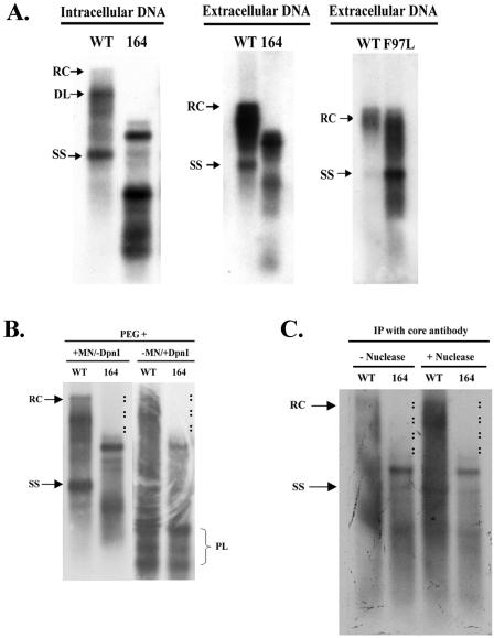
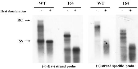
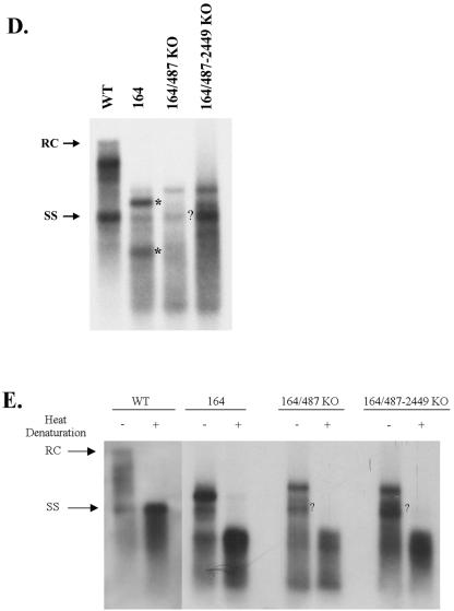
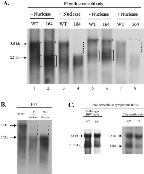
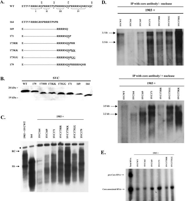
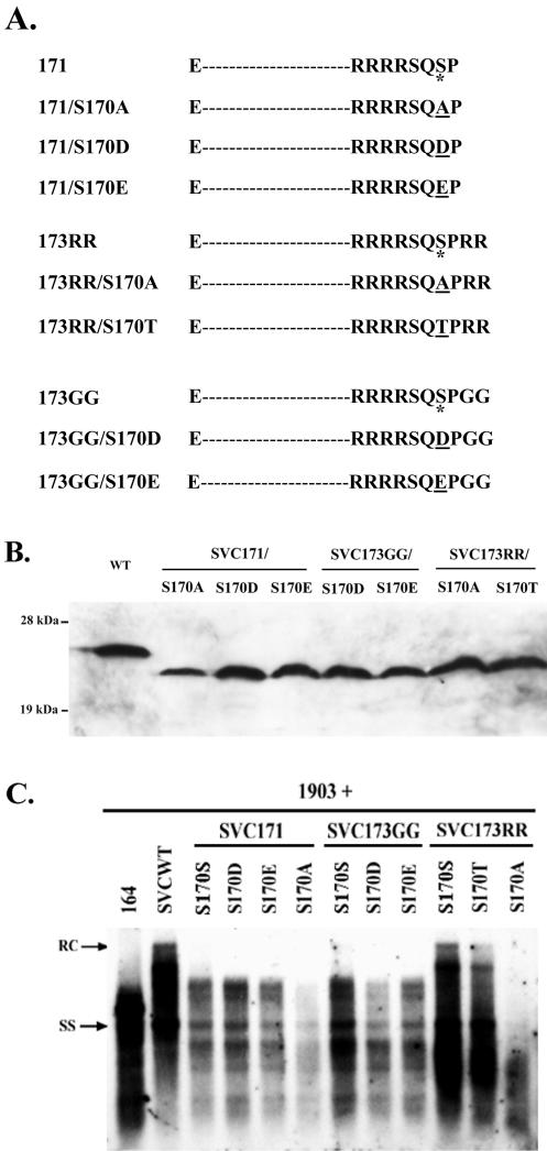
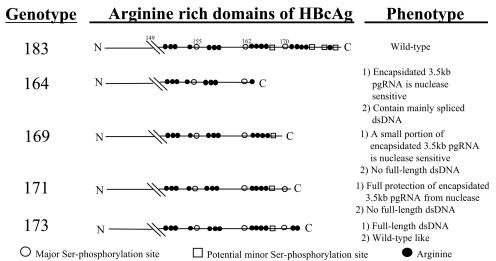
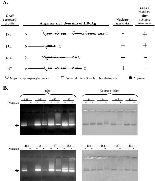
Previously, human hepatitis B virus (HBV) mutant 164, which has a truncation at the C terminus of the HBV core antigen (HBcAg), was speculated to secrete immature genomes. For this study, we further characterized mutant 164 by different approaches. In addition to the 3.5-kb pregenomic RNA (pgRNA), the mutant preferentially encapsidated the 2.2-kb or shorter species of spliced RNA, which can be reverse transcribed into double-stranded DNA before virion secretion. We observed that mutant 164 produced less 2.2-kb spliced RNA than the wild type. Furthermore, it appeared to produce at least two different populations of capsids: one encapsidated a nuclease-sensitive 3.5-kb pgRNA while the other encapsidated a nuclease-resistant 2.2-kb spliced RNA. In contrast, the wild-type core-associated RNA appeared to be resistant to nuclease. When arginines and serines were systematically restored at the truncated C terminus, the core-associated DNA and nuclease-resistant RNA gradually increased in both size and signal intensity. Full protection of encapsidated pgRNA from nuclease was observed for HBcAg 1-171. A full-length positive-strand DNA phenotype requires positive charges at amino acids 172 and 173. Phosphorylation at serine 170 is required for optimal RNA encapsidation and a full-length positive-strand DNA phenotype. RNAs encapsidated in Escherichia coli by capsids of HBcAg 154, 164, and 167, but not HBcAg 183, exhibited nuclease sensitivity; however, capsid instability after nuclease treatment was observed only for HBcAg 164 and 167. A new hypothesis is proposed here to highlight the importance of a balanced charge density for capsid stability and intracapsid anchoring of RNA templates.


Figures

FIG. 1.
FIG. 1.
(A) Different intracellular and extracellular HBV DNA patterns between arginine-rich domain-defective mutant 164 and WT HBV. The replicative intermediates for both mutant 164 and the WT (left), extracted from the PEG-precipitated intracellular core particles at 5 days posttransfection, were analyzed by Southern blotting with a 3.1-kb HBV DNA probe. An analysis of viral secretion (middle) was performed on the cesium chloride gradient fractions corresponding to virion particles (pooled from fractions 10, 12, 14, and 16; density = 1.23 g/ml). A lambda HindIII size marker was included (data not shown). The full-length relaxed circle form (RC) of DNA at the 4.0-kb position, the full-length double-stranded linear form (DL) of DNA at the 3.2-kb position, and the full-length single-stranded form (SS) of DNA at the 1.5-kb position are indicated by arrows. The immature secretion phenotype of a naturally occurring HBV capsid variant (F97L) was included for comparison (right) (58). (B) Comparison of Southern patterns of genomic DNAs of WT HBV and mutant 164 isolated by the PEG method with or without micrococcal nuclease (MN) and DpnI treatment (59). The missing DNA signals in mutant 164 are highlighted by adjacent dots. PL, three major DpnI-digested fragments of transfected plasmid DNA. (C) Comparison of Southern patterns of genomic DNAs of wild-type HBV and mutant 164 isolated by the immunoprecipitation (IP) method with or without nuclease treatment (see Materials and Methods). The missing DNA signals in mutant 164 are highlighted by adjacent dots.
FIG. 2.
FIG. 2.
Heat denaturation and a positive-strand-specific probe demonstrated that PEG-precipitated intracellular mutant 164 core particles contained shorter single-stranded and double-stranded DNA replicative intermediates. Replicative intermediates were extracted from intracellular core particles at 5 days posttransfection and were heat denatured (5 min at 100°C) before gel electrophoresis. Southern blot analysis was performed with a double-stranded HBV DNA probe as described in the legend to Fig. 1 (left). The double-stranded HBV DNA probe was then removed, and the same nitrocellulose filter was rehybridized with a positive-strand-specific riboprobe (right). The minus-strand probe was not used because it gives virtually identical Southern patterns as the double-stranded probe.
FIG. 3.
FIG. 3.
PCR cloning and sequencing analysis of viral DNAs present in mutant 164 virions. (A) Oligonucleotide primers used to PCR amplify virus particle-associated DNAs. Nucleotide positions are given according to the ayw subtype (12). (B) Nested PCR products were loaded in a 1.2% agarose gel and detected by ethidium bromide staining. M1 and M2, DNA molecular weight markers (λ DNA/HindIII and φX174 RF DNA/HaeIII, respectively); NC, negative control (H2O); PC, positive control (plasmid 164). (C) Amplified DNAs from mutant 164 within the 0.5- to 1.3-kb (a) and 0.2- to 0.4-kb (b) ranges were cloned into a TA cloning vector, and colonies were screened by sequencing. The cartoon summarizes all of the spliced DNAs obtained from the PCR products. Nucleotide positions at the intron-exon boundaries are given according to the ayw subtype (12). ɛ, packaging signal; SD, splice donor; SA, splice acceptor. (D) Mutations at the major splice acceptor site alone (164/487KO) or at both the major splice donor and major splice acceptor sites (164/487-2449KO) eliminated strong signals (*) presumably corresponding to the ssDNA and dsDNA replicative intermediates of mutant 164. The question mark indicates DNA signals of splice mutants which happened to comigrate with the ssDNA of WT HBV. (E) Heat denaturation experiments demonstrated that no ssDNAs of splice mutants (164/487KO and 164/487-2449KO) were really of the same size as the ssDNA of WT HBV.
FIG. 3.
FIG. 3.
PCR cloning and sequencing analysis of viral DNAs present in mutant 164 virions. (A) Oligonucleotide primers used to PCR amplify virus particle-associated DNAs. Nucleotide positions are given according to the ayw subtype (12). (B) Nested PCR products were loaded in a 1.2% agarose gel and detected by ethidium bromide staining. M1 and M2, DNA molecular weight markers (λ DNA/HindIII and φX174 RF DNA/HaeIII, respectively); NC, negative control (H2O); PC, positive control (plasmid 164). (C) Amplified DNAs from mutant 164 within the 0.5- to 1.3-kb (a) and 0.2- to 0.4-kb (b) ranges were cloned into a TA cloning vector, and colonies were screened by sequencing. The cartoon summarizes all of the spliced DNAs obtained from the PCR products. Nucleotide positions at the intron-exon boundaries are given according to the ayw subtype (12). ɛ, packaging signal; SD, splice donor; SA, splice acceptor. (D) Mutations at the major splice acceptor site alone (164/487KO) or at both the major splice donor and major splice acceptor sites (164/487-2449KO) eliminated strong signals (*) presumably corresponding to the ssDNA and dsDNA replicative intermediates of mutant 164. The question mark indicates DNA signals of splice mutants which happened to comigrate with the ssDNA of WT HBV. (E) Heat denaturation experiments demonstrated that no ssDNAs of splice mutants (164/487KO and 164/487-2449KO) were really of the same size as the ssDNA of WT HBV.
FIG. 4.
FIG. 4.
(A) Duplicate experiments with two independent transfections and Northern blot analyses of viral RNAs present in immunoprecipitated core particles of mutant 164 and wild-type HBV, with or without nuclease treatment. The intracellular core particles of mutant 164 encapsidated more 2.2-kb and lower molecular weight RNAs than did wild-type HBV. Upon nuclease treatment, RNAs between the 2.2- and 3.5-kb positions disappeared from mutant 164. *, putative encapsidated 3.5-kb pgRNA; brackets, 2.2-kb RNA region before nuclease treatment; dotted areas, nuclease-sensitive core particle-associated RNAs between 2.2 and 3.5 kb. (B) Comparison by Northern blotting of encapsidated viral RNAs of mutant 164 isolated by two different methods of capsid preparation, i.e., PEG and immunoprecipitation. Dotted areas, nuclease-sensitive core particle-associated RNAs between 2.2 and 3.5 kb. (C) Northern blot analysis of total intracellular RNAs by use of a core-specific probe (right) or a full-length 3.2-kb vector-free HBV DNA probe (left). The 3.5-kb core-specific RNA was used here as an internal reference for quantification of the 2.2-kb spliced RNA in the right panel. Briefly, total RNAs were isolated at 3 days posttransfection, and 30 μg was loaded into each lane. The 3.5-kb pgRNA and the 2.2-kb spliced RNA species are indicated by arrows. The envelope-specific nonspliced 2.3- and 2.1-kb subgenomic RNA species did not cross-hybridize with the core-specific probe.
FIG. 5.
FIG. 5.
Functional analysis of the C-terminal domain of the core protein by restoring amino acids to mutant 164. (A) C-terminal amino acid sequences of the various truncated core proteins used for this study. The four arginine (R)-rich domains (I to IV) as well as the phosphorylation sites (*) at serine residues 155, 162, and 170 are indicated. Missense mutations are underlined. (B) Western blot analysis of truncated core proteins by use of a rabbit anticore antibody. The weaker signal obtained for SVC169 was not representative. (C) Wild-type and mutant core proteins were supplied in trans to core-defective HBV replicon 1903 (58). Their respective complementation effects on intracellular HBV DNA replication were analyzed by Southern blotting. The dot represents incomplete nuclease digestion of the input plasmid during the HBV DNA extraction from PEG-precipitated capsids. All samples were run in the same gel, but the exposure time for autoradiography was shorter for the first two lanes. (D) Northern blot analysis of immunoprecipitated core-associated RNAs (see the legend to Fig. 4) of the various mutants shown in panel A. trans-Complementation experiments were conducted as described for panel C. Top, no nuclease treatment; bottom, nuclease treatment. (E) Primer extension analysis of 5′ ends of core-associated RNAs, including both 3.5- and 2.2-kb RNA species. No precore RNA was encapsidated. As a control, the cytoplasmic total RNA was used in the primer extension assay. The identities of the bands corresponding to precore and core RNAs were based on an adjacent DNA sequencing ladder (data not shown).
FIG. 6.
FIG. 6.
Analysis of potential phosphorylation effect at serine residue 170 in various HBcAg contexts (HBcAg 1-171, HBcAg 1-173RR, and HBcAg 1-173GG). (A) C-terminal amino acid sequences of Ser-170 mutants used for this study. Missense mutations are underlined. (B) A Western blot analysis of truncated core proteins by use of an anticore antibody revealed no major difference in expression levels. (C) Wild-type and mutant core proteins were supplied in trans to the core-deficient and replication-defective HBV mutant 1903 (58). Their respective complementation effects on intracellular HBV DNA replication were analyzed by Southern blotting. (D) Primer extension analysis of 5′ ends of core-associated RNAs. The PEG method with nuclease treatment was used for capsid preparation.
FIG. 6.
FIG. 6.
Analysis of potential phosphorylation effect at serine residue 170 in various HBcAg contexts (HBcAg 1-171, HBcAg 1-173RR, and HBcAg 1-173GG). (A) C-terminal amino acid sequences of Ser-170 mutants used for this study. Missense mutations are underlined. (B) A Western blot analysis of truncated core proteins by use of an anticore antibody revealed no major difference in expression levels. (C) Wild-type and mutant core proteins were supplied in trans to the core-deficient and replication-defective HBV mutant 1903 (58). Their respective complementation effects on intracellular HBV DNA replication were analyzed by Southern blotting. (D) Primer extension analysis of 5′ ends of core-associated RNAs. The PEG method with nuclease treatment was used for capsid preparation.
FIG. 7.
FIG. 7.
Cartoon summary of arginine-rich domain mutants of HBcAg and their respective phenotypes. Open circles, major serine phosphorylation sites at amino acids 155, 162, and 170; solid circles, arginine; open squares, potential minor serine phosphorylation sites.
FIG. 8.
FIG. 8.
(A) Illustration of C-terminally truncated HBcAg capsids 154, 164, 167, and 183 (full length). See the legend to Fig. 7 for an explanation of the symbols. The results from the nuclease assays shown in panel B are also summarized here. (B) Nuclease sensitivity assays of E. coli-derived C-terminally truncated HBcAg capsids 154, 164, 167, and 183 via native agarose gel electrophoresis. Twenty micrograms of purified capsid was treated with micrococcal nuclease for 1 h (top) or 3 h (bottom). The treated and untreated control capsid preparations were run in 1% agarose gels containing EtBr to measure their nucleic acid contents. The same gels were subsequently stained with Coomassie blue to visualize the capsid proteins. Arrows indicate the T=4 icosahedral HBcAg capsids (32). Fainter bands above the arrows include contaminating larger particles (unpublished results). Note the substantial loss of EtBr staining upon nuclease treatment of HBcAg 154, 164, and 167 capsids and the little or no loss with the HBcAg 183 capsid. An apparent reduction in the amount of capsid protein was also observed for capsids 164 and 167, but not for capsid 154 or 183. The loss of staining or the diffuse staining pattern by Coomassie blue for capsids 164 and 167 on agarose gels was likely due to a drastic conformational change or disintegration of the icosahedral capsids.

Similar articles

Cited by

References

    1. Bartenschlager, R., M. Junker-Niepmann, and H. Schaller. 1990. The P gene product of hepatitis B virus is required as a structural component for genomic RNA encapsidation. J. Virol. 64:5324-5332. - PMC - PubMed
    1. Beames, B., and R. E. Lanford. 1993. Carboxy-terminal truncations of the HBV core protein affect capsid formation and the apparent size of encapsidated HBV RNA. Virology 194:597-607. - PubMed
    1. Berkowitz, R., A. Ohagen, S. Hoglund, and S. P. Goff. 1995. Retroviral nucleocapsid domains mediate the specific recognition of genomic viral RNAs by chimeric Gag polyproteins during RNA packaging in vivo. J. Virol. 69:6445-6456. - PMC - PubMed
    1. Birnbaum, F., and M. Nassal. 1990. Hepatitis B virus nucleocapsid assembly: primary structure requirements in the core protein. J. Virol. 64:3319-3330. - PMC - PubMed
    1. Bruss, V., and D. Ganem. 1991. The role of envelope proteins in hepatitis B virus assembly. Proc. Natl. Acad. Sci. USA 88:1059-1063. - PMC - PubMed

Publication types

LinkOut - more resources