

doi: 10.1128/JVI.79.3.1911-1917.2005.
Enhanced cytotoxicity without internuclear spread of adenovirus upon cell fusion by measles virus glycoproteins
Affiliations
- PMID: 15650215
- PMCID: PMC544120
- DOI: 10.1128/JVI.79.3.1911-1917.2005
Item in Clipboard
Enhanced cytotoxicity without internuclear spread of adenovirus upon cell fusion by measles virus glycoproteins
J Virol.
2005 Feb.
Abstract
The efficiency of viruses in cancer therapy is enhanced by proteins that mediate the fusion of infected cells with their neighbors. It was reported that replication-competent adenovirus particles can spread between nuclei within fusion-generated syncytia. To assess this conjecture, we generated fusogenic adenoviruses that express a balanced ratio of the F and H glycoproteins of measles virus. The viruses displayed enhanced cytotoxicity but largely unchanged replication efficiencies compared to a nonfusogenic virus. Most notably, the virus genomes did not spread through fusion-generated multinuclear cells. Hence, adenovirus replication in syncytia remains largely restricted to initially transduced nuclei.
Figures
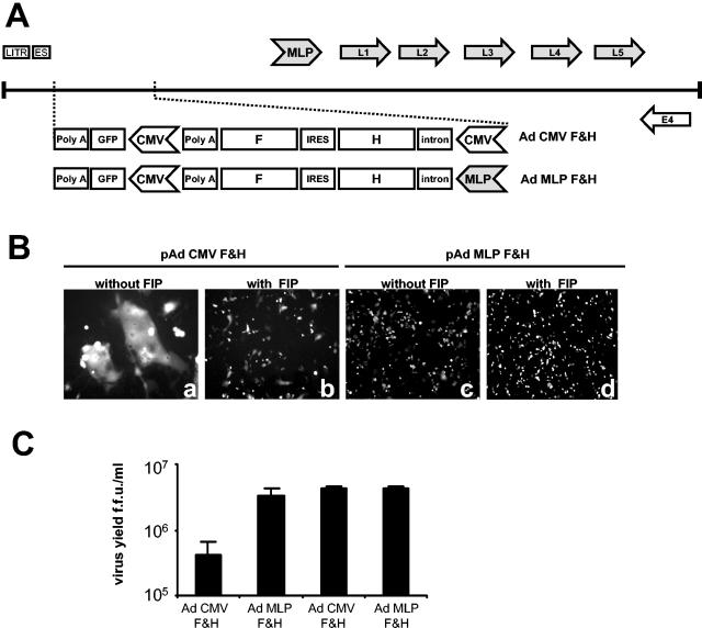
Improving the yield of recombinant fusogenic adenovirus by use of FIP or by late expression of fusogenic proteins. (A) Schematic presentation of fusogenic adenoviruses (linear genomes). Vector genomes are depicted and indicate the assembly of the transgene. The virus genome starts with the left inverted terminal repeat region (LITR) and the encapsidation signal (ES). The positions of some of the early (E) and late (L) genes are indicated. The promoters used to express F and H were either the cytomegalovirus major immediate early promoter (CMV) or the adenovirus major late promoter (MLP). The genes encoding the H and F genes of measles virus (strain Edmonston) were separated by the IRES of encephalomyocarditis virus. A polyadenylation signal (polyA) was then followed by a CMV promoter-driven expression cassette for GFP. (B) Cell morphology upon transfection of GFP-expressing virus genomes. HER911 cells were transfected (Lipofectamine Plus; Invitrogen) with linearized plasmids containing the virus genomes shown in panel A. FIP was added as indicated. After 48 h, green fluorescence was monitored by microscopy. (C) Virus yield 10 days after transfection, given in fluorescence-forming units (f.f.u.) per ml of cell lysate. HER911 cells were transfected and treated as described for panel B. After 10 days, the emerged virus was harvested and quantified on a fresh monolayer of HER911 cells as described previously (23, 40). The average titers of at least three independent experiments are shown along with standard errors. The left two columns reflect results obtained in the absence of FIP; the right two columns show the titers obtained in the presence of FIP.
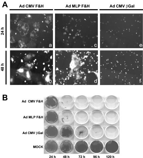
Cytotoxicity induced by fusogenic adenovirus. (A) Timing of fusion. HER911 cells were infected with the indicated virus preparations at an MOI of 0.01 for the indicated times, followed by monitoring of the distribution of GFP by fluorescence microscopy. (B) Time course of cytotoxic effects. HER911 cells were transduced with the indicated viruses (MOI = 1) for the indicated times. Subsequently, detached cells were washed off the plate, and the remaining cell monolayer was stained with crystal violet.
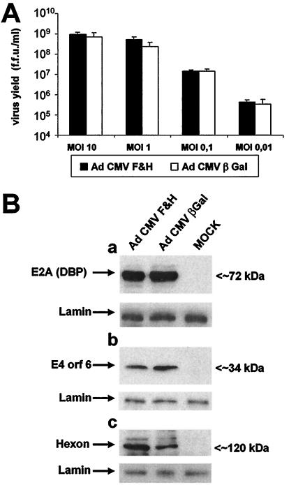
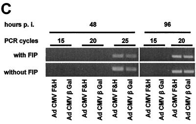
Replication yield of fusogenic adenovirus. (A) Replication in the presence or absence of fusion. HER911 cells were infected with the indicated virus preparations. At 48 h postinfection, the viruses were harvested and titrated. (B) HER911 cells were infected as indicated (MOI = 0.1). The expression of the viral gene products E2A, E4orf6, and hexon was monitored by immunoblotting 48 h after infection. Lamin was stained as an input control. (C) Upon infection as described for panel B, the amounts of viral DNA were determined at the indicated times postinfection by semiquantitative PCRs with the indicated numbers of PCR cycles in the presence or absence of FIP. The PCR products were visualized by ethidium bromide staining after agarose gel electrophoresis.
Replication yield of fusogenic adenovirus. (A) Replication in the presence or absence of fusion. HER911 cells were infected with the indicated virus preparations. At 48 h postinfection, the viruses were harvested and titrated. (B) HER911 cells were infected as indicated (MOI = 0.1). The expression of the viral gene products E2A, E4orf6, and hexon was monitored by immunoblotting 48 h after infection. Lamin was stained as an input control. (C) Upon infection as described for panel B, the amounts of viral DNA were determined at the indicated times postinfection by semiquantitative PCRs with the indicated numbers of PCR cycles in the presence or absence of FIP. The PCR products were visualized by ethidium bromide staining after agarose gel electrophoresis.
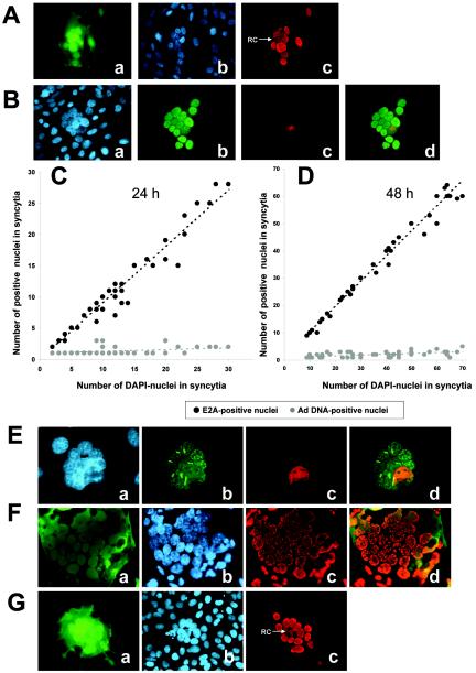
Formation of replication centers within syncytia. HER911 cells were infected at an MOI of 0.01 with Ad CMV F&H. To avoid extracellular spread, we added an antibody against the fiber knob 3 h after infection. After 48 h, immunostaining of the E2A 72-kDa DNA-binding protein was performed. (A) GFP distribution (a) and DAPI staining (to visualize all nuclei) (b) were monitored, and E2A was visualized by immunofluorescence (c). The typical distribution of E2A in replication centers (RC) within one of the nuclei is indicated by an arrow. (B) The total DNA was stained with DAPI (a), and E2A-72 kDa was visualized by immunofluorescence (b). In addition, viral DNA was stained by FISH (c). Superimposed patterns of E2A and viral DNA are shown in panel d. At least 30 syncytia were evaluated at 24 (C) or 48 (D) h postinfection by FISH and immunostaining. The total number of nuclei within each syncytium was plotted against the number of E2A-positive nuclei (black dots) and against the number of cell nuclei that stained positive for viral DNA (gray dots). (E) To monitor hexon assembly, we transduced HER911 cells as described above and then added an antibody against the fiber knob. After 48 h, immunostaining of the hexon protein was performed (b). The cells were also stained with DAPI to detect all nuclei (a), and FISH was employed to detect viral DNA (c). The patterns of hexon and viral DNA were superimposed (d). (F) To study the formation of replication centers in preformed syncytia, we transfected 911 cells with an expression plasmid for the measles virus F and H proteins. After 24 h, syncytia had formed, and further fusion was stopped by the addition of FIP. The cells were then infected with the wild-type adenovirus dl309, followed by incubation for another 24 h. E2A was then stained as described for panel A. (G) U2OS cells seeded at a low density were coinfected with wild-type adenovirus dl309 and Ad CMV F&H at an MOI of 10. Three hours later, fresh U2OS cells were added to form a confluent monolayer, and an antibody against the fiber knob was added. Forty-eight hours later, the cells were fixed and E2A was stained as described for panel A.
Similar articles
-
Importance of the cytoplasmic tails of the measles virus glycoproteins for fusogenic activity and the generation of recombinant measles viruses.J Virol. 2002 Jul;76(14):7174-86. doi: 10.1128/jvi.76.14.7174-7186.2002. J Virol. 2002. PMID: 12072517 Free PMC article.
-
Measles virus: both the haemagglutinin and fusion glycoproteins are required for fusion.J Gen Virol. 1991 Feb;72 ( Pt 2):439-42. doi: 10.1099/0022-1317-72-2-439. J Gen Virol. 1991. PMID: 1993882
-
Use of viral fusogenic membrane glycoproteins as novel therapeutic transgenes in gliomas.Hum Gene Ther. 2001 May 1;12(7):811-21. doi: 10.1089/104303401750148766. Hum Gene Ther. 2001. PMID: 11339897
-
Virus-Mediated Cell-Cell Fusion.Int J Mol Sci. 2020 Dec 17;21(24):9644. doi: 10.3390/ijms21249644. Int J Mol Sci. 2020. PMID: 33348900 Free PMC article. Review.
-
Nuclear organization of replication and gene expression in adenovirus-infected cells.Curr Top Microbiol Immunol. 1995;199 ( Pt 1):99-117. doi: 10.1007/978-3-642-79496-4_7. Curr Top Microbiol Immunol. 1995. PMID: 7555063 Review. No abstract available.
Cited by
-
Use of cell fusion proteins to enhance adenoviral vector efficacy as an anti-cancer therapeutic.Cancer Gene Ther. 2021 Aug;28(7-8):745-756. doi: 10.1038/s41417-020-0192-9. Epub 2020 Jul 1. Cancer Gene Ther. 2021. PMID: 32606392 Review.
-
Principles of Virus Uncoating: Cues and the Snooker Ball.Traffic. 2016 Jun;17(6):569-92. doi: 10.1111/tra.12387. Epub 2016 Mar 31. Traffic. 2016. PMID: 26875443 Free PMC article. Review.
-
Syncytia formation affects the yield and cytotoxicity of an adenovirus expressing a fusogenic glycoprotein at a late stage of replication.Gene Ther. 2008 Sep;15(17):1240-5. doi: 10.1038/gt.2008.94. Epub 2008 May 29. Gene Ther. 2008. PMID: 18509378 Free PMC article.
-
In situ tumor vaccination with adenovirus vectors encoding measles virus fusogenic membrane proteins and cytokines.World J Gastroenterol. 2007 Jun 14;13(22):3063-70. doi: 10.3748/wjg.v13.i22.3063. World J Gastroenterol. 2007. PMID: 17589921 Free PMC article.
References
-
- Ahmed, A., D. Jevremovic, K. Suzuki, T. Kottke, J. Thompson, S. Emery, K. Harrington, A. Bateman, and R. Vile. 2003. Intratumoral expression of a fusogenic membrane glycoprotein enhances the efficacy of replicating adenovirus therapy. Gene Ther. 10:1663-1671. - PubMed
-
- Alemany, R., C. Balague, and D. T. Curiel. 2000. Replicative adenoviruses for cancer therapy. Nat. Biotechnol. 18:723-727. - PubMed
-
- Bateman, A., F. Bullough, S. Murphy, L. Emiliusen, D. Lavillette, F. L. Cosset, R. Cattaneo, S. J. Russell, and R. G. Vile. 2000. Fusogenic membrane glycoproteins as a novel class of genes for the local and immune-mediated control of tumor growth. Cancer Res. 60:1492-1497. - PubMed
-
- Bischoff, J. R., D. H. Kirn, A. Williams, C. Heise, S. Horn, M. Muna, L. Ng, J. A. Nye, A. Sampson-Johannes, A. Fattaey, and F. McCormick. 1996. An adenovirus mutant that replicates selectively in p53-deficient human tumor cells. Science 274:373-376. - PubMed
-
- Bucheit, A. D., S. Kumar, D. M. Grote, Y. Lin, V. von Messling, R. B. Cattaneo, and A. K. Fielding. 2003. An oncolytic measles virus engineered to enter cells through the CD20 antigen. Mol. Ther. 7:62-72. - PubMed
Publication types
MeSH terms
Substances
Grants and funding
LinkOut - more resources
Full Text Sources
