

Attenuating mutations of the matrix gene of influenza A/WSN/33 virus
- PMID: 15650216
- PMCID: PMC544140
- DOI: 10.1128/JVI.79.3.1918-1923.2005
Attenuating mutations of the matrix gene of influenza A/WSN/33 virus
Abstract
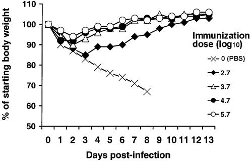
The matrix protein (M1) of influenza virus plays an essential role in viral replication. Our previous studies have shown that basic amino acids 101RKLKR105 of M1 are involved in RNP binding and nuclear localization. For the present work, the functions of 101RKLKR105 were studied by introducing mutations into the M gene of influenza virus A/WSN/33 by reverse genetic methods. Individual substitution, R101S or R105S, had a minimal effect on viral replication. In contrast, the double mutation R101S-R105S was synergistic and resulted in temperature sensitivity reflected by reduced viral replication at a restrictive temperature. To investigate the in vivo effect on infection, BALB/c mice were infected with either A/WSN/33 wild-type (Wt) or mutant viruses and assessed for signs of illness, viral replication in the lungs, and survival rates. The results from mouse studies indicated that the R101S-R105S double mutant virus was strongly attenuated, while single mutant viruses R101S and R105S were minimally attenuated compared to A/WSN33 Wt under the same conditions. In challenge studies, mice immunized by infection with R101S-R105S were fully protected from lethal challenge with A/WSN/33. The replication and attenuating properties of R101S-R105S suggest its potential in development of live influenza virus vaccines.
Figures
Similar articles
-
Introduction of a temperature-sensitive phenotype into influenza A/WSN/33 virus by altering the basic amino acid domain of influenza virus matrix protein.J Virol. 2004 Sep;78(18):9585-91. doi: 10.1128/JVI.78.18.9585-9591.2004. J Virol. 2004. PMID: 15331690 Free PMC article.
-
A live attenuated H1N1 M1 mutant provides broad cross-protection against influenza A viruses, including highly pathogenic A/Vietnam/1203/2004, in mice.J Infect Dis. 2009 Dec 15;200(12):1874-83. doi: 10.1086/648405. J Infect Dis. 2009. PMID: 19909080
-
Synergism of co-mutation of two amino acid residues in NS1 protein increases the pathogenicity of influenza virus in mice.Virus Res. 2010 Aug;151(2):200-4. doi: 10.1016/j.virusres.2010.05.007. Epub 2010 May 28. Virus Res. 2010. PMID: 20546807
-
A new approach to an influenza life vaccine: haemagglutinin cleavage site mutants generated by reverse genetics.Berl Munch Tierarztl Wochenschr. 2006 Mar-Apr;119(3-4):186-91. Berl Munch Tierarztl Wochenschr. 2006. PMID: 16573209 Review.
-
Structured model of influenza virus replication in MDCK cells.Biotechnol Bioeng. 2004 Oct 5;88(1):1-14. doi: 10.1002/bit.20096. Biotechnol Bioeng. 2004. PMID: 15384040 Review.
Cited by
-
Genetic contributions to influenza virus attenuation in the rat brain.J Neurovirol. 2008 Apr;14(2):136-42. doi: 10.1080/13550280701885563. J Neurovirol. 2008. PMID: 18444085
-
The compensatory G88R change is essential in restoring the normal functions of influenza A/WSN/33 virus matrix protein 1 with a disrupted nuclear localization signal.J Virol. 2013 Jan;87(1):345-53. doi: 10.1128/JVI.02024-12. Epub 2012 Oct 17. J Virol. 2013. PMID: 23077315 Free PMC article.
-
Matrix protein 1: A comparative in silico study on different strains of influenza A H5N1 Virus.Bioinformation. 2006 Nov 22;1(7):253-6. doi: 10.6026/97320630001253. Bioinformation. 2006. PMID: 17597902 Free PMC article.
-
Influenza A virus inhibits alveolar fluid clearance in BALB/c mice.Am J Respir Crit Care Med. 2008 Nov 1;178(9):969-76. doi: 10.1164/rccm.200803-455OC. Epub 2008 Aug 8. Am J Respir Crit Care Med. 2008. PMID: 18689466 Free PMC article.
-
Mutations in influenza virus M1 CCHH, the putative zinc finger motif, cause attenuation in mice and protect mice against lethal influenza virus infection.J Virol. 2006 Jun;80(12):5697-707. doi: 10.1128/JVI.02729-05. J Virol. 2006. PMID: 16731908 Free PMC article.
References
-
- Cox, N. J., F. Kitame, A. P. Kendal, H. F. Maassab, and C. Naeve. 1988. Identification of sequence changes in the cold-adapted, live attenuated influenza vaccine strain, A/Ann Arbor/6/60 (H2N2). Virology 167:554-567. - PubMed
MeSH terms
Substances
LinkOut - more resources
Full Text Sources
Other Literature Sources
Medical
