. 2005 Feb;115(2):459-67.
doi: 10.1172/JCI22462.

MyD88-dependent induction of allergic Th2 responses to intranasal antigen

Affiliations

MyD88-dependent induction of allergic Th2 responses to intranasal antigen

Damani A Piggott et al. J Clin Invest. 2005 Feb.

Abstract

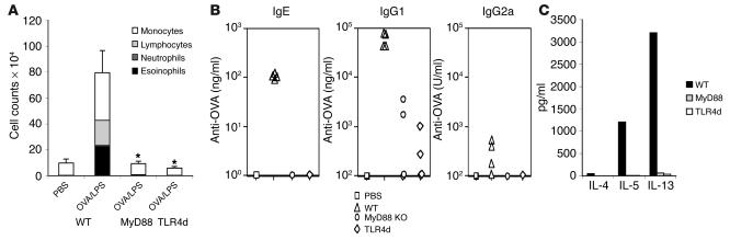
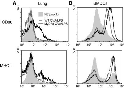
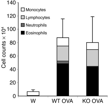
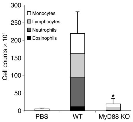
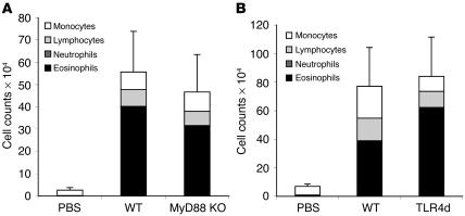
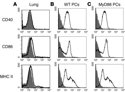
MyD88 is a common Toll-like receptor (TLR) adaptor molecule found to be essential for induction of adaptive Th1 immunity. Conversely, innate control of adaptive Th2 immunity has been shown to occur in a MyD88-independent manner. In this study, we show that MyD88 is an essential innate component in the induction of TLR4-dependent Th2 responses to intranasal antigen; thus we demonstrate what we believe to be a novel role for MyD88 in pulmonary Th2 immunity. Induction of the MyD88-independent type I IFN response to LPS is defective in the pulmonary environment. Moreover, in the absence of MyD88, LPS-induced upregulation of costimulatory molecule expression on pulmonary DCs is defective, in contrast to what has been observed with bone marrow-derived DCs (BMDCs). Reconstitution of Th2 responses occurs upon adoptive pulmonary transfer of activated BMDCs to MyD88-deficient recipients. Furthermore, the dependence of Th2 responses on MyD88 is governed by the initial route of antigen exposure; this demonstrates what we believe are novel site-specific innate mechanisms for control of adaptive Th2 immunity.


Figures

Figure 1
Figure 1
Th2 responses to intranasal protein antigen are MyD88 dependent. (A) BAL inflammatory cells of WT, MyD88-deficient, or TLR4d mice sensitized intranasally with OVA/LPS and WT mice sensitized with PBS. Exposed mice were then challenged 2 weeks later with inhaled OVA. Total bar height represents total BAL cell number, and error bars are based on total cell numbers. Stacked bars represent cell differential counts. n = 4. *P < 0.05, total BAL cell number from WT vs. MyD88-deficient and WT vs. TLR4d. One representative experiment of 4 is shown. (B) OVA-specific serum antibody responses by ELISA after challenge with OVA in WT, MyD88-deficient, and TLR4d mice sensitized as described in A. P < 0.05, WT vs. MyD88-deficient and WT vs. TLR4d, for IgE and IgG1 responses. (C) Cytokine production from lung draining lymph nodes after challenge with OVA in WT, MyD88-deficient, and TLR4d mice sensitized as described in A.
Figure 2
Figure 2
Th2-mediated lung inflammation can occur in the absence of MyD88. (A) BAL inflammatory cells of WT and MyD88-deficient mice sensitized with OVA/LPS intranasally or OVA/alum i.p. Exposed mice were then challenged 2 weeks later with inhaled OVA. Total bar height represents total BAL cell number at day 21 after challenge. Error bars are based on total cell numbers. Stacked bars represent cell differential counts. n = 4. *P < 0.05, WT vs. MyD88-deficient mice primed with OVA/LPS. One representative experiment of 3 is shown. (B) BAL inflammatory cells of WT and MyD88-deficient mice sensitized intranasally with OVA/LPS or OVA/TNF-α. Exposed mice were then challenged 2 weeks later with inhaled OVA. Total bar height represents total BAL cell number at day 21 after challenge. Error bars are based on total cell numbers. Stacked bars represent cell differential counts. n = 4. *P < 0.05, WT vs. MyD88-deficient mice primed with OVA/LPS. One representative experiment of 3 is shown.
Figure 3
Figure 3
Phenotype of lung cells in the absence of collagenase digestion. (A) Unstimulated lung cells harvested from WT mice with and without collagenase digestion and analyzed by FACS. Percentage yield and total number of CD11c+ cells obtained from resting lung by collagenase digestion (D) are shown compared with those from nondigested tissue (N). (B) Analysis of non-CD11c+ cell population from nondigested lung tissue by FACS. Expression of cell markers Mac1, B220, Gr1, γδ, NK, CD4, and CD8 on non-CD11c+ cells is shown on forward scatter (Fsc) compared with an isotype control. Numbers indicate the percentage of live cells positive for the indicated cell marker.
Figure 4
Figure 4
Pulmonary DCs are not activated by LPS in the absence of MyD88 in vivo. (A) MHC class II and CD86 FACS analysis, at 48 hours, of CD11c+ lung cells from WT and MyD88-deficient mice sensitized to OVA/LPS or PBS intranasally. (B) MHC class II and CD86 FACS analysis of CD11c+ BMDCs generated from WT and MyD88-deficient mice pulsed with OVA/LPS overnight or left untreated (no Tx).
Figure 5
Figure 5
MyD88-deficient pulmonary DCs demonstrate defective activation in response to LPS in vitro. (A) Change in CD86 expression at 24 hours on CD11c+ lung cells from WT mice and CD11c+ BMDCs generated from WT mice responding to stimulation with varying doses of LPS in vitro. Bar heights represent the mean of 3 independent experiments. (B) Change in CD86 expression at 24 hours on CD11c+ BMDCs generated from WT and MyD88-deficient mice. (C) Change in CD86 expression at 24 hours on CD11c+ lung cells from WT and MyD88-deficient mice. *P < 0.05, WT vs. MyD88-deficient lung cells and WT lung cells vs. WT BMDCs.
Figure 6
Figure 6
IFN- β is not induced in response to LPS exposure in the lung. WT mice were exposed to PBS, LPS, and CpG. Lung cells were harvested 18 hours after exposure and lysed, and RNA was extracted. Five micrograms of each RNA sample was analyzed by RNase protection assay using a RiboQuant mCK-3b probe. Fold induction of (A) TNF-α and (B) IFN-β is shown relative to PBS.
Figure 7
Figure 7
Mature BMDCs can reconstitute Th2 responses in MyD88-deficient mice. BAL inflammatory cells of WT and MyD88-deficient mice given OVA/LPS–pulsed BMDCs intranasally and challenged intranasally with OVA after 2 weeks. Total bar height represents total BAL cell number at day 21. Error bars are based on total cell numbers. Stacked bars represent cell differential counts. n = 4. W, unpulsed WT BMDCs to WT recipient; WT OVA, OVA/LPS–pulsed WT BMDCs to WT recipient; KO OVA, OVA/LPS–pulsed WT BMDCs to MyD88-deficient recipient. P = 0.82, WT OVA vs. KO OVA. One representative experiment of 3 is shown.
Figure 8
Figure 8
High-dose LPS response is MyD88 dependent. BAL inflammatory cells of WT and MyD88-deficient mice sensitized with OVA/high-dose LPS intranasally. Exposed mice were challenged 2 weeks later with inhaled OVA. Total bar height represents total BAL cell number at day 21 after challenge. Error bars are based on total cell numbers. Stacked bars represent cell differential counts. *P < 0.05, OVA/high-dose LPS–primed WT vs. MyD88-deficient mice. One representative experiment of 3 is shown.
Figure 9
Figure 9
Th2 responses to i.p. protein antigen are TLR4 and MyD88 independent. BAL inflammatory cells of WT mice and MyD88-deficient mice (A) or TLR4d mice (B) sensitized i.p. with OVA/LPS. Exposed mice were challenged 2 weeks later with inhaled OVA. Total bar height represents total BAL cell number at day 21 after challenge. Error bars are based on total cell numbers. Stacked bars represent cell differential counts. n = 4. P = 0.68, WT vs. TLR4d; P = 0.33, WT vs. MyD88-deficient.
Figure 10
Figure 10
Pulmonary APCs demonstrate low maturational status compared with peritoneal APCs. Unstimulated lung cells harvested from WT mice (A) and unstimulated peritoneal cells (PCs) from WT mice (B) and MyD88-deficient mice (C), analyzed by FACS. MHC class II, CD86, and CD40 expression on live cells is shown compared with isotype control (shaded).

References

    1. Constant SL, Bottomly K. Induction of Th1 and Th2 CD4+ T cell responses: the alternative approaches. Annu. Rev. Immunol. 1997;15:297–322. - PubMed
    1. Eisenbarth SC, Piggott DA, Bottomly K. The master regulators of allergic inflammation: dendritic cells in Th2 sensitization. Curr. Opin. Immunol. 2003;15:620–626. - PubMed
    1. Lenschow DJ, Walunas TL, Bluestone JA. CD28/B7 system of T cell costimulation. Annu. Rev. Immunol. 1996;14:233–258. - PubMed
    1. Sharpe AH, Freeman GJ. The B7-CD28 superfamily. Nat. Rev. Immunol. 2002;2:116–126. - PubMed
    1. Janeway CA, Jr, Medzhitov R. Innate immune recognition. Annu. Rev. Immunol. 2002;20:197–216. - PubMed

Publication types

MeSH terms