

2-aminoethoxydiphenyl borate stimulates pulmonary C neurons via the activation of TRPV channels
- PMID: 15653710
- PMCID: PMC1783973
- DOI: 10.1152/ajplung.00439.2004
2-aminoethoxydiphenyl borate stimulates pulmonary C neurons via the activation of TRPV channels
Abstract
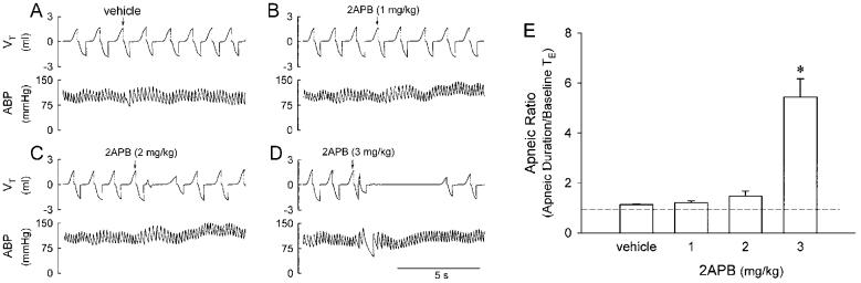
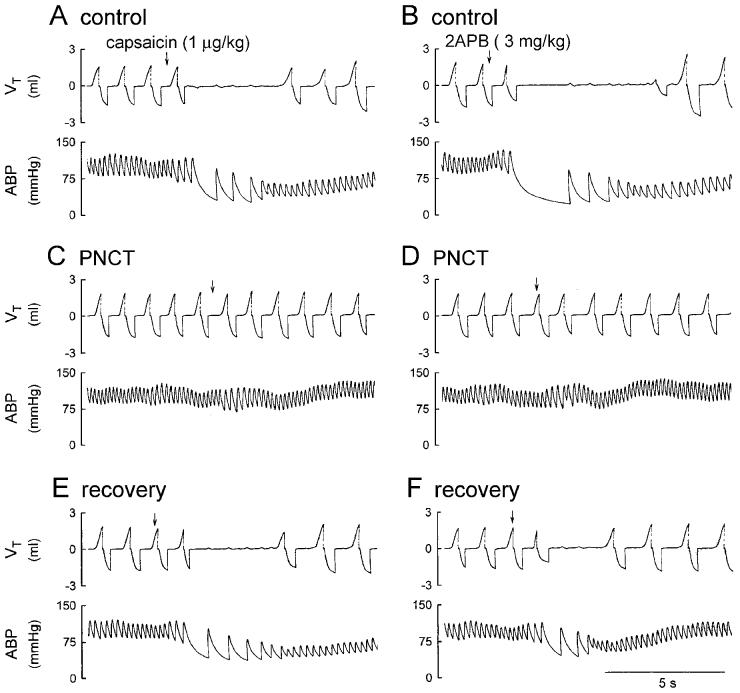
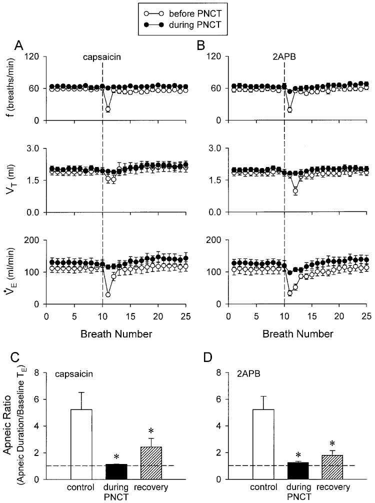
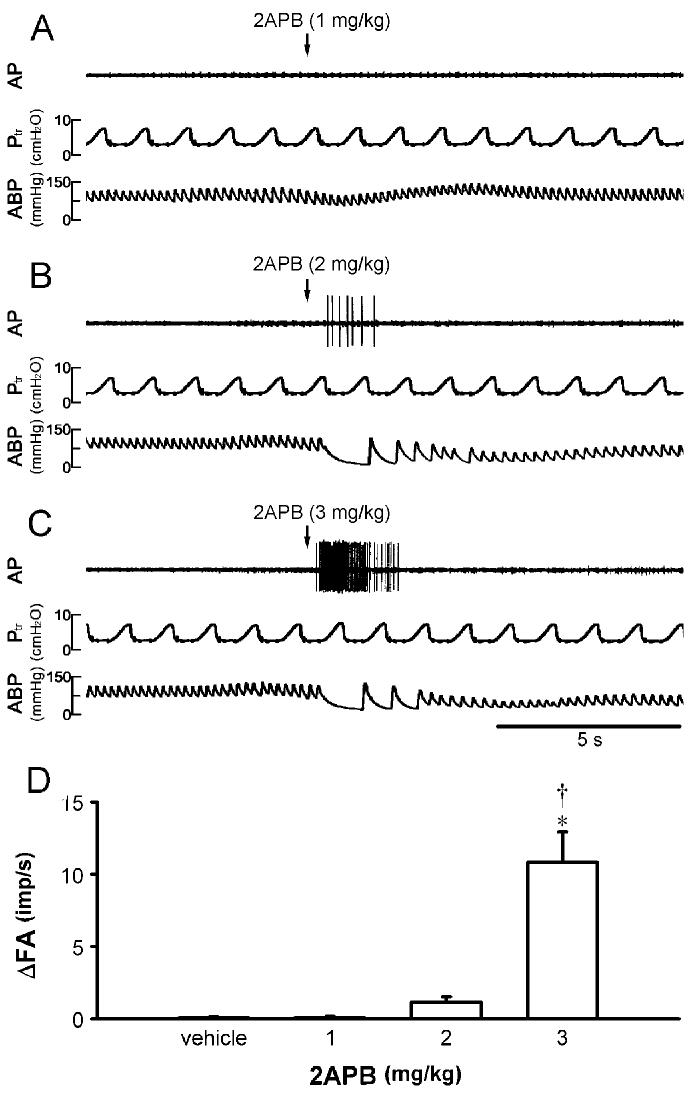
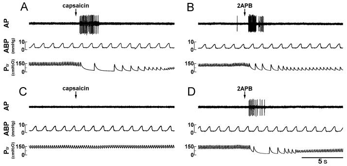
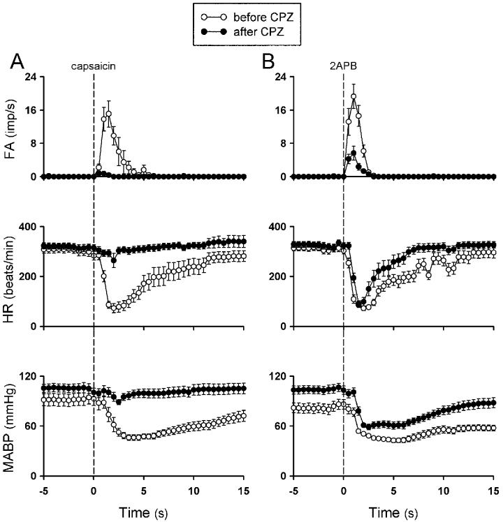
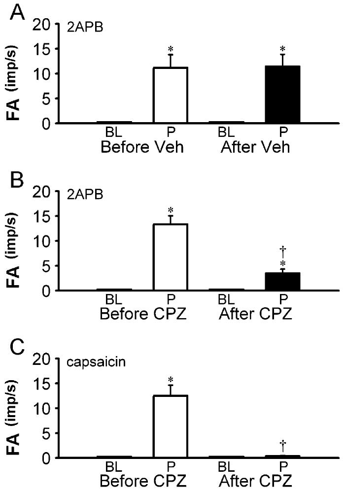
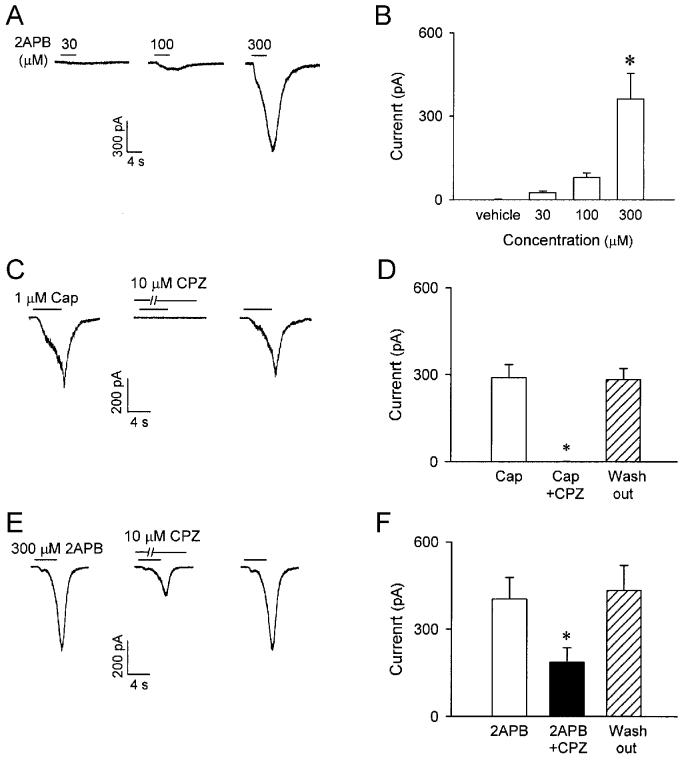
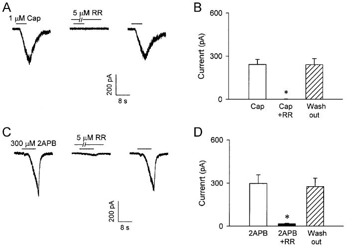
This study was carried out to determine the effect of 2-aminoethoxydiphenyl borate (2-APB), a common activator of transient receptor potential vanilloid (TRPV) type 1, 2, and 3 channels, on cardiorespiratory reflexes, pulmonary C fiber afferents, and isolated pulmonary capsaicin-sensitive neurons. In anesthetized, spontaneously breathing rats, intravenous bolus injection of 2-APB elicited the pulmonary chemoreflex responses, characterized by apnea, bradycardia, and hypotension. After perineural treatment of both cervical vagi with capsaicin to block the conduction of C fibers, 2-APB no longer evoked any of these reflex responses. In open-chest and artificially ventilated rats, 2-APB evoked an abrupt and intense discharge in vagal pulmonary C fibers in a dose-dependent manner. The stimulation of C fibers by 2-APB was attenuated but not abolished by capsazepine, a selective antagonist of the TRPV1, which completely blocked the response to capsaicin in these C fiber afferents. In isolated pulmonary capsaicin-sensitive neurons, 2-APB concentration dependently evoked an inward current that was partially inhibited by capsazepine but almost completely abolished by ruthenium red, an effective blocker of all TRPV channels. In conclusion, 2-APB evokes a consistent and distinct stimulatory effect on pulmonary C fibers in vivo and on isolated pulmonary capsaicin-sensitive neurons in vitro. These results establish the functional evidence demonstrating that TRPV1, V2, and V3 channels are expressed on these sensory neurons and their terminals.
Figures
Similar articles
-
Stimulation of pulmonary vagal C-fibres by anandamide in anaesthetized rats: role of vanilloid type 1 receptors.J Physiol. 2002 Mar 15;539(Pt 3):947-55. doi: 10.1113/jphysiol.2001.013290. J Physiol. 2002. PMID: 11897863 Free PMC article.
-
Effect of increasing temperature on TRPV1-mediated responses in isolated rat pulmonary sensory neurons.Am J Physiol Lung Cell Mol Physiol. 2008 Mar;294(3):L563-71. doi: 10.1152/ajplung.00336.2007. Epub 2008 Jan 4. Am J Physiol Lung Cell Mol Physiol. 2008. PMID: 18178674
-
Effect of olvanil and anandamide on vagal C-fiber subtypes in guinea pig lung.Br J Pharmacol. 2005 Oct;146(4):596-603. doi: 10.1038/sj.bjp.0706339. Br J Pharmacol. 2005. PMID: 16056239 Free PMC article.
-
Functional morphology and physiological properties of bronchopulmonary C-fiber afferents.Anat Rec A Discov Mol Cell Evol Biol. 2003 Jan;270(1):17-24. doi: 10.1002/ar.a.10005. Anat Rec A Discov Mol Cell Evol Biol. 2003. PMID: 12494486 Review.
-
Interaction between TRPA1 and TRPV1: Synergy on pulmonary sensory nerves.Pulm Pharmacol Ther. 2015 Dec;35:87-93. doi: 10.1016/j.pupt.2015.08.003. Epub 2015 Aug 14. Pulm Pharmacol Ther. 2015. PMID: 26283426 Free PMC article. Review.
Cited by
-
Hypoxia augments TRPM3-mediated calcium influx in vagal sensory neurons.Auton Neurosci. 2023 Jul;247:103095. doi: 10.1016/j.autneu.2023.103095. Epub 2023 Apr 29. Auton Neurosci. 2023. PMID: 37146443 Free PMC article.
-
Transient receptor potential vanilloid channels in hypertension, inflammation, and end organ damage: an imminent target of therapy for cardiovascular disease?Curr Opin Cardiol. 2008 Jul;23(4):356-63. doi: 10.1097/HCO.0b013e32830460ad. Curr Opin Cardiol. 2008. PMID: 18520720 Free PMC article. Review.
-
Calcium transient evoked by TRPV1 activators is enhanced by tumor necrosis factor-{alpha} in rat pulmonary sensory neurons.Am J Physiol Lung Cell Mol Physiol. 2010 Oct;299(4):L483-92. doi: 10.1152/ajplung.00111.2010. Epub 2010 Jul 16. Am J Physiol Lung Cell Mol Physiol. 2010. PMID: 20639352 Free PMC article.
-
Activation of TRPV4 Regulates Respiration through Indirect Activation of Bronchopulmonary Sensory Neurons.Front Physiol. 2016 Feb 29;7:65. doi: 10.3389/fphys.2016.00065. eCollection 2016. Front Physiol. 2016. PMID: 26973533 Free PMC article.
-
A novel native store-operated calcium channel encoded by Orai3: selective requirement of Orai3 versus Orai1 in estrogen receptor-positive versus estrogen receptor-negative breast cancer cells.J Biol Chem. 2010 Jun 18;285(25):19173-83. doi: 10.1074/jbc.M110.102582. Epub 2010 Apr 15. J Biol Chem. 2010. PMID: 20395295 Free PMC article.
References
-
- Adriaensen D, Timmermans JP, Brouns I, Berthoud HR, Neuhuber WL, Scheuermann DW. Pulmonary intraepithelial vagal nodose afferent nerve terminals are confined to neuroepithelial bodies: an anterograde tracing and confocal microscopy study in adult rats. Cell Tissue Res. 1998;293:395–405. - PubMed
-
- Baluk P, Nadel JA, McDonald DM. Substance P-immunoreactive sensory axons in the rat respiratory tract: a quantitative study of their distribution and role in neurogenic inflammation. J Comp Neurol. 1992;319:586–598. - PubMed
-
- Benham CD, Davis JB, Randall AD. Vanilloid and TRP channels: a family of lipid-gated cation channels. Neuropharmacology. 2002;42:873–888. - PubMed
-
- Bootman MD, Collins TJ, Mackenzie L, Roderick HL, Berridge MJ, Peppiatt CM. 2-aminoethoxydiphenyl borate (2-APB) is a reliable blocker of store-operated Ca2+ entry but an inconsistent inhibitor of InsP3-induced Ca2+ release. FASEB J. 2002;16:1145–1150. - PubMed
Publication types
MeSH terms
Substances
Grants and funding
LinkOut - more resources
Full Text Sources
Other Literature Sources
Molecular Biology Databases