

Multilayer structures in lipid monolayer films containing surfactant protein C: effects of cholesterol and POPE
- PMID: 15653721
- PMCID: PMC1305360
- DOI: 10.1529/biophysj.104.050823
Multilayer structures in lipid monolayer films containing surfactant protein C: effects of cholesterol and POPE
Abstract
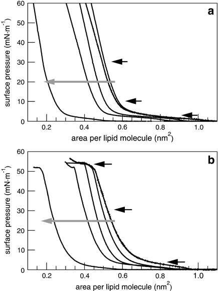
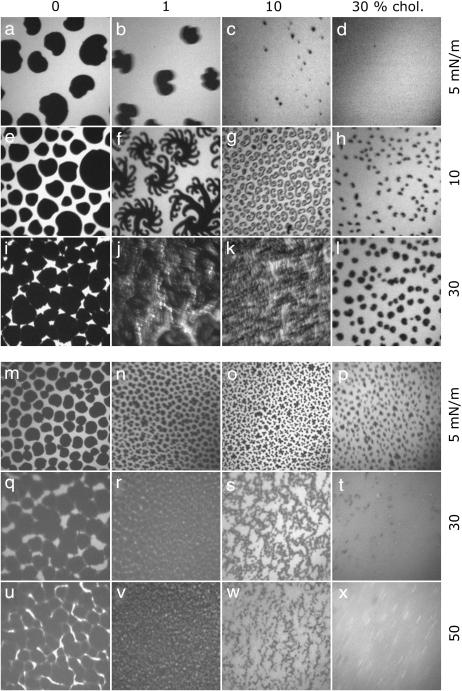
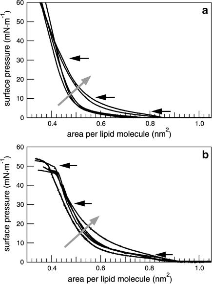
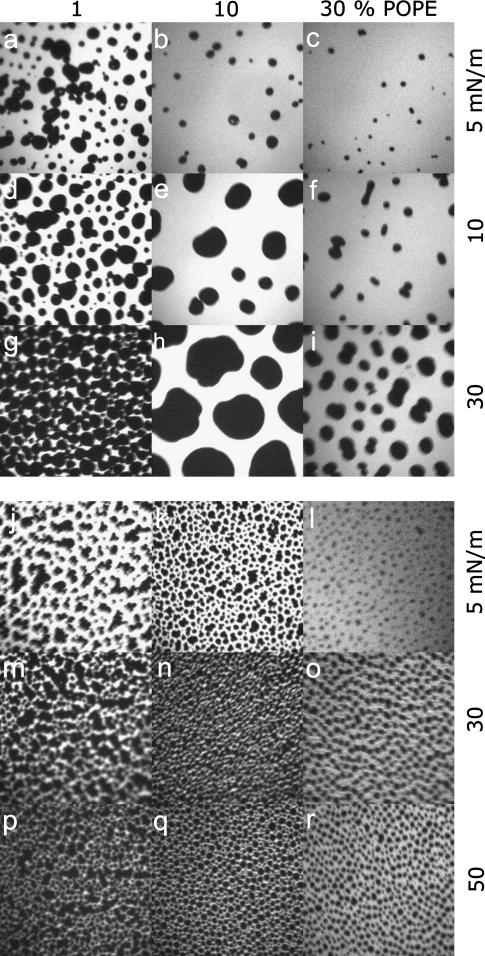
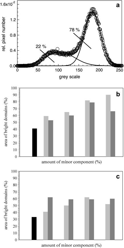
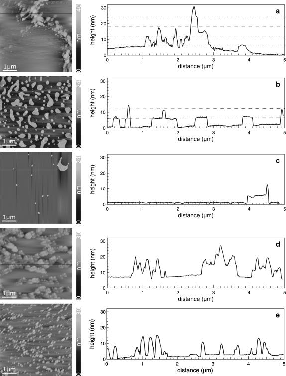
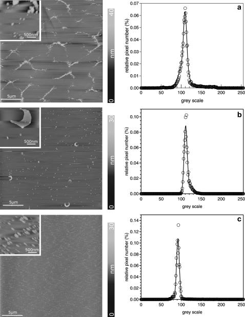
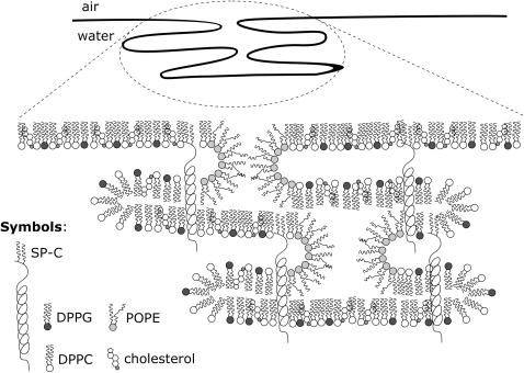
The influence of cholesterol and POPE on lung surfactant model systems consisting of DPPC/DPPG (80:20) and DPPC/DPPG/surfactant protein C (80:20:0.4) has been investigated. Cholesterol leads to a condensation of the monolayers, whereas the isotherms of model lung surfactant films containing POPE exhibit a slight expansion combined with an increased compressibility at medium surface pressure (10-30 mN/m). An increasing amount of liquid-expanded domains can be visualized by means of fluorescence light microscopy in lung surfactant monolayers after addition of either cholesterol or POPE. At surface pressures of 50 mN/m, protrusions are formed which differ in size and shape as a function of the content of cholesterol or POPE, but only if SP-C is present. Low amounts of cholesterol (10 mol %) lead to an increasing number of protrusions, which also grow in size. This is interpreted as a stabilizing effect of cholesterol on bilayers formed underneath the monolayer. Extreme amounts of cholesterol (30 mol %), however, cause an increased monolayer rigidity, thus preventing reversible multilayer formation. In contrast, POPE, as a nonbilayer lipid thought to stabilize the edges of protrusions, leads to more narrow protrusions. The lateral extension of the protrusions is thereby more influenced than their height.
Figures
Similar articles
-
Effects of cholesterol on surface activity and surface topography of spread surfactant films.Biochemistry. 2002 Dec 17;41(50):15007-16. doi: 10.1021/bi0256532. Biochemistry. 2002. PMID: 12475250
-
Pulmonary surfactant protein C containing lipid films at the air-water interface as a model for the surface of lung alveoli.Mol Membr Biol. 1995 Jan-Mar;12(1):93-9. doi: 10.3109/09687689509038502. Mol Membr Biol. 1995. PMID: 7767391
-
Formation of three-dimensional protein-lipid aggregates in monolayer films induced by surfactant protein B.Biophys J. 2000 Aug;79(2):904-18. doi: 10.1016/S0006-3495(00)76346-6. Biophys J. 2000. PMID: 10920022 Free PMC article.
-
Protein-lipid interactions and surface activity in the pulmonary surfactant system.Chem Phys Lipids. 2006 Jun;141(1-2):105-18. doi: 10.1016/j.chemphyslip.2006.02.017. Epub 2006 Mar 20. Chem Phys Lipids. 2006. PMID: 16600200 Review.
-
Lipid-protein interactions of hydrophobic proteins SP-B and SP-C in lung surfactant assembly and dynamics.Pediatr Pathol Mol Med. 2001 Nov-Dec;20(6):445-69. doi: 10.1080/pdp.20.6.445.469. Pediatr Pathol Mol Med. 2001. PMID: 11699574 Review.
Cited by
-
An elevated level of cholesterol impairs self-assembly of pulmonary surfactant into a functional film.Biophys J. 2007 Jul 15;93(2):674-83. doi: 10.1529/biophysj.107.106310. Epub 2007 May 4. Biophys J. 2007. PMID: 17483162 Free PMC article.
-
Influence of lipid saturation grade and headgroup charge: a refined lung surfactant adsorption model.Biophys J. 2008 Jul;95(2):699-709. doi: 10.1529/biophysj.108.131102. Epub 2008 Apr 4. Biophys J. 2008. PMID: 18390619 Free PMC article.
-
Effects of palmitoylation on dynamics and phospholipid-bilayer-perturbing properties of the N-terminal segment of pulmonary surfactant protein SP-C as shown by 2H-NMR.Biophys J. 2008 Sep;95(5):2308-17. doi: 10.1529/biophysj.108.132845. Epub 2008 May 23. Biophys J. 2008. PMID: 18502795 Free PMC article.
-
Influence of Cholesterol on the Insertion and Interaction of SARS-CoV-2 Proteins with Lipid Membranes.ACS Appl Bio Mater. 2025 Jun 16;8(6):5380-5394. doi: 10.1021/acsabm.5c00776. Epub 2025 Jun 6. ACS Appl Bio Mater. 2025. PMID: 40474679 Free PMC article.
-
Nanoparticle interaction with model lung surfactant monolayers.J R Soc Interface. 2010 Feb 6;7 Suppl 1(Suppl 1):S15-26. doi: 10.1098/rsif.2009.0329.focus. Epub 2009 Oct 21. J R Soc Interface. 2010. PMID: 19846443 Free PMC article.
References
-
- Ahn, T., and C. H. Yun. 1999. Phase properties of liquid-crystalline phosphatidylcholine/phosphatidylethanolamine bilayers revealed by fluorescent probes. Arch. Biochem. Biophys. 369:288–294. - PubMed
-
- Akinbi, H. T., J. S. Breslin, M. Ikegami, H. S. Iwamoto, J. C. Clark, J. A. Whitsett, A. H. Jobe, and T. E. Weaver. 1997. Rescue of SP-B knockout mice with a truncated SP-B proprotein. Function of the C-terminal propeptide. J. Biol. Chem. 272:9640–9647. - PubMed
-
- Amrein, M., A. von Nahmen, and M. Sieber. 1997. A scanning force- and fluorescence light microscopy study of the structure and function of a model pulmonary surfactant. Eur. Biophys. J. 26:349–357. - PubMed
-
- Burns, A. R. 2003. Domain structure in model membrane bilayers investigated by simultaneous atomic force microscopy and fluorescence imaging. Langmuir. 19:8358–8363.
Publication types
MeSH terms
Substances
LinkOut - more resources
Full Text Sources
Medical
